Viagra gibt es mittlerweile nicht nur als Original, sondern auch in Form von Generika. Diese enthalten denselben Wirkstoff Sildenafil. Patienten suchen deshalb nach viagra generika schweiz, um ein günstigeres Präparat zu finden. Unterschiede bestehen oft nur in Verpackung und Preis.
Piers.org
Progress In Electromagnetics Research Symposium Proceedings, Moscow, Russia, August 19–23, 2012 145
X-ray Image Processing in Studying Jawbone Tissues
J. Mikulka and M. Kabrda
Department of Theoretical and Experimental Electrical Engineering
Brno University of Technology, Kolejn´ı 4, Brno 612 00, Czech Republic
Abstract— Image processing in biomedical applications is strongly developing issue. Manymethods and approaches for image preprocessing, segmentation and visualization were described.
This paper describes X-ray image processing. The aim of processing is to segment regions ofjawbone cysts and evaluate their local descriptors. It is necessary to choose suitable segmentationmethod because of adverse parameters of regions. The regions of the cysts are of low contrastand the pixel intensity distribution is not homogenous. The semiautomatic live-wire methodwas chosen. This approach to image segmentation is faster and more accurate than manualsegmentation. It is very good compromise between simple manual edge tracing and automaticmethods such as thresholding, watershed segmentation or other methods whose results mustbe post-processed. The second step of processing is to evaluate local descriptors of segmentedregions which correspond to cysts. Several parameters were chosen to describe these regions —region area, mean gray value of intensities, modal gray value of intensities, standard deviationof intensities, minimal and maximal gray value of intensities, integrated intensity, median ofintensities and shape descriptors of region (perimeter, circularity, aspect ratio, roundness andsolidity). Values of these parameters will be used in following development of semiautomaticprocessing method with regard to current assessment of cysts by doctors.
Cysts are defined as pathological cavities with their own capsule, epithelium and liquid or mushycontent. The cyst wall is formed by fibrous tissue. A typical property of the cyst is its expansivegrowth. The cyst wall functions as a semipermeable membrane, permeable in one direction for thesurrounding liquid. The result is a gradual enlargement of the cyst [1].
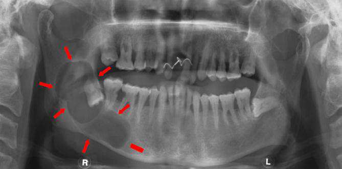
Follicular cysts (Fig. 1) arise from the epithelium of tooth germ and develop either between a
naked crown and the joined inner and outer epithelium or enamel or between the two layers ofepithelium. Their appearance can be triggered by a primary disorder in the development of toothgerms. Follicular cysts can occur individually or multiply [2]. The majority of follicular cysts mostfrequently appear in the region of lower jawbone. Bulky cysts can lead to face-deforming swelling.
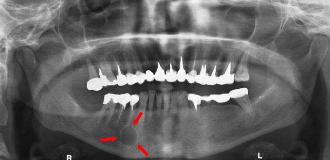
Radicular cysts (Fig. 1) belong to the most frequently occurring jawbone cysts. They are believedto appear due to inflammatory excitation of Malasser epithelium remnants of periodontal creviceby the infected content of the root duct of a transversely situated tooth. Cysts are associated withteeth without pulp and they occur in all age categories (mostly 30 and 40 years of age), and are abit more frequent in males. The follicle of radicular cyst is usually of round or oval shape [1].
To obtain images of the afflicted jawbones, X-ray examination is very often performed in practice
using the orthopantomograph, which enables panoramic images of whole jawbones to be obtained.
The paper describes the first steps in proposing methods for the processing of OPG images and
their implementation in the automation of the assessment of the two types of cyst. The aim of theproposal is to make and train a model from actual medical data and then to use it for an automaticclassification of cysts according to the existing scale used in manual processing.
2. IMAGE PROCESSING CHAIN
The image processing chain currently consists of orthopantomograph detection, segmentation ofthe cystic region, and description of regions in terms of local properties. The existing solution will
Figure 1: OPG image of follicular cyst on the left and radicular cyst on the right.
PIERS Proceedings, Moscow, Russia, August 19–23, 2012
later be complemented with the results of a classification based on the physician's knowledge which,together with a selection of local descriptors will be sufficient to train a suitably selected model(Fig. 2). The trained model will subsequently be used for automated computer-aided classificationof cysts.
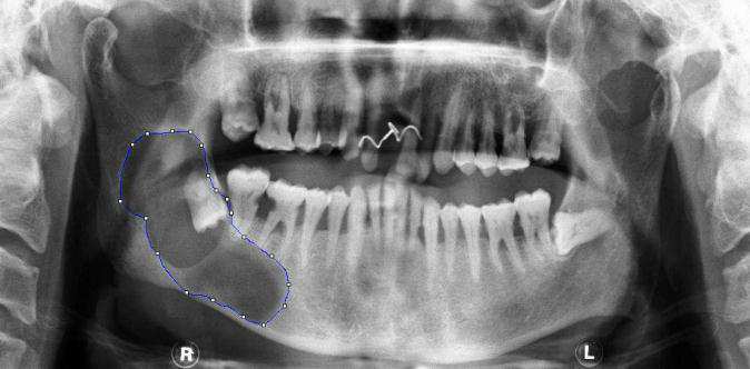
In view of the properties of the regions that represent the jawbone cysts, and the necessity of
delineating precisely the given region in order to obtain corresponding local descriptors, the live-wire semi-automatic segmentation method was chosen. The method enables fast manual delineationof the region of interest, using the shortest path algorithm. The image first undergoes convolutionusing the Sobel filter [6], by means of which the edges in the image are enhanced. The user marksout individual points in the image, between which the optimum shortest path is sought, whichwill run along the image edges [7]. In our case the shortest path is being sought using the Djikstralgorithm. The algorithm is finite (for any finite input the algorithm will end) because in eachcycle run exactly one node is added to the set of visited nodes. Thus there are at most as manycycle runs as there are peaks in the graph. Let us imagine we have a graph G, in which we wantto find the shortest path. Let us determine that V is a set containing all the peaks of graph Gand E is a set of all edges of graph G. The principle of the algorithm consist in that for each peakv from V the algorithm remembers the shortest length of path that needs to be taken to get tothe peak. Let us denote this value d[v]. In the beginning, all the peaks have the value d[v] = ∞,excepting the initial peak s, the corresponding value of which is d[s] = 0. Infinity expresses anunknown path to the peak. The algorithm further utilizes the sets Z and N, where N contains thehitherto unvisited and Z the visited peaks. The algorithm is repeated cyclically until the set Nis empty. In each algorithm cycle one peak vmin is added from N into Z. It is the peak with the
least value d[v] from all the peaks v from N. For each peak u, with one edge from vmin leading to
it (its length expressed as l(vmin, u)) the following algorithm is applied: if (d[vmin] + l[vmin]) < d[u],
then the value d[vmin] + l[vmin, u] is assigned to d[u]. If this condition is not satisfied, nothing will
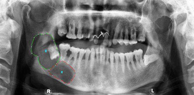
happen. When the algorithm ends, then for each peak v from the set V the length of its shortestpath from the initial peak s will be assigned to d[v] [8]. The live-wire method is suitable for thesegmentation of images in which the region of interest is in individual images always in a differentposition and always has a different shape. This method is thus in our case the most suitable forimage segmentation. One or more regions can be marked/delineated simultaneously in the image(Fig. 3).
The aim of further processing the segmented images is to obtain local descriptors of regions that
represent jawbone cysts. The Image J program was used to evaluate statistically the distributionof pixel intensities in cystic regions and to establish the shape parameters of these regions. In thisphase of research the following parameters were evaluated in the submitted set of images: area,
Figure 2: Image processing chain designed for automated classification of follicular and radicular cysts.
Figure 3: Semiautomatic segmentation using the live-wire method — one delineated region, several delineatedregions.
Progress In Electromagnetics Research Symposium Proceedings, Moscow, Russia, August 19–23, 2012 147
mean intensity value in the region, standard deviation of pixel intensities in the region, median ofpixel intensities in the region, modal value of pixel intensities in the region, minimum and maximumvalues of pixel intensities in the region, and integral of pixel intensities in the region. Also, the shapeparameters of segmented regions were established: perimeter of the region, circularity accordingto (1), ratio of the major and minor half-exes of the inscribed ellipse, roundness according to (2),and convexity according to the relation (3):
C = 4 · π ·
R = 4 ·
π · a2major
where C is the circularity of the region, S is the area of the region, P is the perimeter of theregion, R is the roundness of the region, αmajor is the length of the major half-axis of ellipse
inscribed in the region, X is the convexity of the region, and SC is the area of the convex region
described/circumscribed over the region under examination.
3. RESULTS OF PROCESSING
The whole of available database of images, i.e., 13 images with follicular cysts and 18 images withradicular cysts was evaluated using the methodology described. The difference is obvious betweenfollicular and radicular cysts as mentioned in the introductory paragraph. While the follicularcysts are rather of an oblong shape, the radicular cysts are almost always of circular nature. Thedifferent circularity of the two types of cyst provides the possibility of classifying cysts into twobasic groups:
• follicular cyst — circularity: 0.807 ± 0.149,• radicular cyst — circularity: 0.932 ± 0.086.
To enhance the quality of cyst classification, it would be appropriate to select a greater number
of parameters the probability distributions of which do not overlap in the two types of cyst. Onesuch parameter could, for example, be a parameter giving the ratio of the major and minor half-axislengths of inscribed ellipse:
• follicular cyst — ratio of half-axes: 1.975 ± 0.613,• radicular cyst — ratio of half-axes: 1.514 ± 0.401.
Based on subjective assessment of values, the area can also be selected as a suitable candidate forcyst classifier:
• follicular cyst — area: 58029.38 ± 40023.60,• radicular cyst — area: 30249.54 ± 25048.18.
From the discussion of the values it follows that the automated system can be complemented
with a cyst classification according to the cyst type. The next step will be a classification of thelevel of cystic disorder in jawbone according to a scale currently used by physicians for subjectiveassessment of OPG images. Images assessed by physicians will be correlated with the values givenin the tables, and an appropriate model will be made and trained for cyst classification.
Fundamental research described in the paper concerns the area of processing OPG images of jaw-bones with the aim of classifying follicular and radicular cysts. The classification of cysts can bedivided into two types:
• binary classification of the type of cyst: follicular or radicular,• classification of the level of jawbone affliction with cyst disorder on the currently used scale
PIERS Proceedings, Moscow, Russia, August 19–23, 2012
The results of image processing indicated the possibility of using a combination of several parametersfor a binary classification of cysts (circularity, ratio of half-axes of inscribed ellipse, and area ofcystic region). An algorithm was proposed for the classification of the level of jawbone affliction.
The selection of a model and the process of training will be carried out using the Rapid Minersystem in a follow-up work and after the available database has been processed by physicians.
This work was supported in part by the Grant project GAP102/11/0318, 102/12/1104, projectFEKT-S-11-5/1012 and CZ.1.07.2.3.00.20.0175.
1. Pazdera, J., Fundamentals of Oral and Maxillofacial Surgery, UP Olomouc, 2011.
2. ˇ
Cernochova, P., Diagnostika Retinovan´ych Zub˚
u, Grada Publishing, Praha, 2006.
3. Mikulka, J., E. Gescheidtov´a, K. Bartuˇsek, and Z. Sm´ekal, "Processing of MR slices of tem-
poromandibular disc for 3D visualization," PIERS Online, Vol. 6, No. 3, 204–206, 2010.
4. Bartuˇsek, K., E. Gescheidtov´a, and J. Mikulka, "Data processing in studying biological tissues
using MR imaging techniques," 33th the International Conference on Telecommunications andSignal Processing, 171–175, Asszisztenda Szervezo, Budapeˇst, 2010.
5. Sm´ekal, Z., O. Liberda, K. Bartuˇsek, and J. Mikulka, "Data processing in studying the tem-
poromandibular joint, using MR imaging and sonographic techniques," 16th InternationalConference on Digital Signal Processing (DSP 2009), 100–106, Santorini, Greece, 2009.
6. Hlav´aˇc, V. and M. ˇ
Sonka, Image Processing, Grada, Praha, 1992.
7. Baggio, D. L., GPGPU Based Image Segmentation Livewire Algorithm Implemnetation, Tech-
nological Institute of Aeronautics, Sao Jos´e dos Campos, 2007.
8. Brassard, G. and P. Bratley, Algorithmics, Prentice-Hall, Englewood Cliffs, 1988.
Source: https://piers.org/piersproceedings/download.php?file=cGllcnMyMDEyTW9zY293fDFBN18wMTQ1LnBkZnwxMjAxMzEwNTQyMTc=
Non-toxic tumortherapy (NTT) for advanced colorectal cancer with liver metastasis, a pilot study E.Valstar M.D., M. Sc. Stadhouderslaan 30, 2517 HZ The Hague, Holland Eighty-three patients with livermetastases from colorectal cancer and in many cases also metastases elsewhere consulted me in 1988-1997 for non-toxic tumortherapy (NTT). No selection was made. Two regressions during
balanced diet promotes more than good body function, it is an important way to provide vitality and well-being. A good variety of foods should deliver all the nutrients necessary for Aa healthy body in a practical, delicious and creative way. vzv A healthy and fun diet is important for everybody, but particularly those who experience the stress of modern life and lack of time.


