HM Medical Clinic

Even if Viagra is not needed, it is possible that the doctor will be able to determine the etiology of erectile dysfunction and prescribe appropriate treatmen https://vgraustralia.net it doesn't pay to forget about sexual activeness even at the first sings of malfunction.

Mct-14-0013 2203.2214

Published OnlineFirst July 15, 2014; DOI: 10.1158/1535-7163.MCT-14-0013 Cancer Biology and Signal Transduction Quinacrine Overcomes Resistance to Erlotinib by InhibitingFACT, NF-kB, and Cell-Cycle Progression in Non–Small CellLung Cancer Josephine Kam Tai Dermawan1, Katerina Gurova2, John Pink3, Afshin Dowlati4, Sarmishtha De1,Goutham Narla5, Neelesh Sharma4, and George R. Stark1 Erlotinib is a tyrosine kinase inhibitor approved for the treatment of patients with advanced non–small cell lung cancer (NSCLC). In these patients, erlotinib prolongs survival but its benefit remains modest becausemany tumors express wild-type (wt) EGFR or develop a second-site EGFR mutation. To test drug combinationsthat could improve the efficacy of erlotinib, we combined erlotinib with quinacrine, which inhibits the FACT(facilitates chromatin transcription) complex that is required for NF-kB transcriptional activity. In A549(wtEGFR), H1975 (EGFR-L858R/T790M), and H1993 (MET amplification) NSCLC cells, this drug combinationwas highly synergistic, as quantified by Chou–Talalay combination indices, and slowed xenograft tumorgrowth. At a sub-IC50 but more clinically attainable concentration of erlotinib, quinacrine, alone or incombination with erlotinib, significantly inhibited colony formation and induced cell-cycle arrest andapoptosis. Quinacrine decreased the level of active FACT subunit SSRP1 and suppressed NF-kB–dependentluciferase activity. Knockdown of SSRP1 decreased cell growth and sensitized cells to erlotinib. Moreover,transcriptomic profiling showed that quinacrine or combination treatment significantly affected cell-cycle–related genes that contain binding sites for transcription factors that regulate SSRP1 target genes. As potentialbiomarkers of drug combination efficacy, we identified genes that were more strongly suppressed by thecombination than by single treatment, and whose increased expression predicted poorer survival in patientswith lung adenocarcinoma. This preclinical study shows that quinacrine overcomes erlotinib resistance byinhibiting FACT and cell-cycle progression, and supports a clinical trial testing erlotinib alone versus thiscombination in advanced NSCLC. Mol Cancer Ther; 13(9); 2203–14. 2014 AACR.
a better understanding of the pathogenesis of NSCLC, Metastatic non–small cell lung cancer (NSCLC) is the coupled with high-throughput genomic technologies most common cause of cancer death in the United States.
applied to patient tumor samples, has led to a molecular Cytotoxic chemotherapy has historically been the main- classification of NSCLC (and a new generation of "preci- stay of therapy but is associated with only modest sion" therapies). This paradigm is best illustrated by the improvements in patient survival. Over the past decade, identification of activating mutations in EGFR as driversof lung cancer development and progression and thesubsequent demonstration of the clinical benefit of anti- 1Department of Molecular Genetics, Cleveland Clinic Lerner Research EGFR therapies such as erlotinib (Tarceva), a reversible Institute, Cleveland, Ohio. 2Department of Cell Stress Biology, Roswell tyrosine kinase inhibitor (TKI) of EGFR (1). On the other Park Cancer Institute, Buffalo, New York. 3Division of General Medical hand, the clinical benefit of erlotinib is modest in patients Sciences-Oncology, Case Comprehensive Cancer Center, Cleveland,Ohio. 4Case Comprehensive Cancer Center, University Hospitals Seidman with wild-type (wt) EGFR, particularly in those with Cancer Center, Case Western Reserve University, Cleveland, Ohio.
5 concurrent KRAS mutations (2, 3); in addition, even in Department of Medicine, Institute for Transformative Molecular Medicine, Case Western Reserve University, Cleveland, Ohio.
the initially sensitive EGFR mutation–positive patients,population resistance invariably develops through the Note: Supplementary data for this article are available at Molecular CancerTherapeutics Online (http://mct.aacrjournals.org/).
development of second-site EGFR mutations, for example,T790M (4), activation of alternative receptor tyrosine Corresponding Authors: George R. Stark, Department of MolecularGenetics, Cleveland Clinic Lerner Research Institute/NE20, 9500 Euclid kinases, for example, MET amplification (5), and other Avenue, Cleveland, OH 44195.Phone: 216-444-6062; Fax: 216-444-0512; mechanisms including transformation from non–small E-mail: [email protected]; and Neelesh Sharma, Division of Hematology/ cell to small cell histology (6).
Oncology, University Hospitals Case Medical Center, 11100 Euclid Ave-nue, Cleveland, OH 44106. Phone: 216-844-0363; Fax: 216-844-5234; E- Quinacrine was widely used during World War II as an antimalarial agent. Over the last four decades, it has been used for the treatment of giardiasis, tapeworm infesta- 2014 American Association for Cancer Research.
tions, and connective tissue diseases, for example, lupus Published OnlineFirst July 15, 2014; DOI: 10.1158/1535-7163.MCT-14-0013 Dermawan et al.
erythematosus and rheumatoid arthritis (7, 8). Recently, a resuscitation from frozen stocks, and were maintained in chemical screen identified 9-aminoacridines, including DMEM (A549 and H1975) or RPMI-1640 (H1993) medium quinacrine, as activators of p53 and inhibitors of NF-kB supplemented with 5% FBS. All cells were kept at 37C in a (9, 10). NF-kB regulates the expression of genes encoding humidified atmosphere with 5% CO2. A549 has wtEGFR proinflammatory and antiapoptotic proteins. In contrast and mutant KRAS (G61H), H1975 has the activating with the situation in normal cells, it is usually constitu- EGFRL858R mutation as well as the second site EGFRT790M tively active in tumor cells and plays a key role in mutation, which decreases the affinity of the receptor for promoting tumorigenesis, including resistance to many erlotinib, and H1993 has wtEGFR and MET amplification.
cancer therapies (11–13). Indeed, a recent report showedthat inhibition of NF-kB sensitizes NSCLC cells to erloti- Cell proliferation nib-induced cell death (14). Thus, NF-kB is an attractive Cells were seeded in 96-well plates at 1 to 2  103 per target for cancer therapy (12, 15).
well, allowed to attach overnight, and treated with var- Quinacrine is thought to act by intercalating into DNA ious concentrations of erlotinib, quinacrine, or a combi- through its planar acridine ring, whereas its diaminobutyl nation of both in a 5:1 or 10:1 molar ratio. After 72 hours, side chain extends into the DNA minor groove (8). Recent- cell viability was determined by the MTT assay (17). The ly, it was reported that quinacrine and its derivatives combination index (CI) was assessed by using CalcuSyn suppress NF-kB by causing chromatin trapping of the software (Biosoft; refs. 18, 19).
FACT (facilitates chromatin transcription) complex (10), aheterodimer of the structure-specific recognition protein (SSRP1) and suppressor of Ty 16 (SPT16). The normal Cells were seeded in 6-well plates at 500 per well, function of FACT is to promote reorganization of nucleo- allowed to attach overnight, and treated with erlotinib, somes in front of RNA polymerase II during transcription quinacrine, or the combination in triplicate. Drugs were elongation. However, FACT is often expressed in aggres- replaced every 72 hours. After 14 days, cells were fixed sive, undifferentiated cancers, and neoplastic (but not with 100% methanol and stained with 1% crystal violet.
normal) cell growth depends on FACT activity (16). Chro- Colonies were quantified using the cell counter plugin of matin trapping of FACT results in increased phosphory- the NIH ImageJ software (v.1.48).
lation of p53 by the FACT-associated kinase CK2, andreduced NF-kB–dependent transcription because of the Cell-cycle analysis depletion of free active FACT (10).
Cells were treated with 1 mmol/L erlotinib, 3 or 5 mmol/L To improve the clinical benefit of erlotinib in the treat- quinacrine, or a combination of both for 96 or 120 hours, ment of advanced NSCLC, we investigated whether com- and then fixed with 100% cold ethanol at 20C for 1 hour, bination with quinacrine potentiates the ability of erloti- and stained with 3 mmol/L propidium iodide (PI; Invitro- nib to mediate cell death, and the mechanism underlying gen, #P3566) in the presence of RNase for 15 minutes at the observed synergistic effect in NSCLC cells. As a result room temperature. Cell-cycle distribution was assessed of our findings, we are conducting a phase I/II clinical by FACScan (BD Biosciences) analysis.
trial to test the combination of erlotinib and quinacrine inadvanced or metastatic (stage IIIB/IV) NSCLC patients Analysis of apoptosis who have failed at least one prior platinum-based che- Staining was performed using Annexin V-APC motherapy regimen (NCT01839955).
(eBioscience, #88–8007) in conjunction with PI accordingto the manufacturer's protocol, and was assessed by Materials and Methods FACScan. Apoptosis was validated by PARP cleavage and analyzed by the Western method.
Erlotinib was obtained from Selleck Chemicals (# S1023) and dissolved in DMSO. Quinacrine, from Sigma-Aldrich NF-kB luciferase assay (# Q3251), was dissolved in PBS as a 10 mmol/L stock A549 or H1975 cells were infected with the kB-luciferase solution. Dilutions to the required concentrations were lentiviral construct pLA-NFkB-mCMV-luc-H4-puro (or made in DMEM or RPMI-1640 medium. Mouse monoclo- hygro) and stably selected with puromycin or hygromy- nal SSRP1 antibody (# 609701) was from BioLegend.
cin. This NF-kB reporter lentiviral vector consists of a Rabbit polyclonal PARP antibody (# 9542) was from Cell firefly luciferase reporter gene under the control of a Signaling Technology. Mouse monoclonal b-actin anti- minimal (m)CMV promoter and six NF-kB–responsive body (# A5316) was from Sigma. Goat polyclonal Lamin elements from the immunoglobulin light chain gene B (# sc-6216) and mouse monoclonal GAPDH antibody (ref. 20; kind gift from Dr. Peter Chumakov, Russian (# sc-32233) were from Santa Cruz Biotechnology.
Academy of Sciences, Moscow, Russia). The reporter cellswere then seeded in 96-well plates at 1 to 2  103 per well, allowed to attach overnight, and then treated with drugs The human non–small cell lung adenocarcinoma cell and/or IL1. Cells were then harvested in reporter lysis lines A549, H1975, and H1993 were obtained from ATCC buffer (Promega) and assayed for luciferase activity using and passaged for less than 6 months following receipt or the luciferase assay system (Promega).
Mol Cancer Ther; 13(9) September 2014 Molecular Cancer Therapeutics Published OnlineFirst July 15, 2014; DOI: 10.1158/1535-7163.MCT-14-0013 Combination of Quinacrine and Erlotinib in NSCLC DNA-binding assay normalization, and log2 transformation). Probes were The ability of compounds to alter the mobility of plasmid annotated using the HuGene 2.1 st hg19 probeset anno- DNA was tested by incubating plasmid DNA in Tris-EDTA tation files downloaded from the Affymetrix website. Low buffer (pH 8.0) with 10 mmol/L quinacrine or chloroquine intensity probes (probes whose log2 expression levels in at room temperature for 20 minutes followed by electro- the untreated sample were less than the median expres- phoresis (1% agarose gel, 1.5 V/cm constant for 16 hours).
sion level across all probes) were filtered out. Hierarchical Gels were stained with ethidium bromide (0.5 mg/mL) and clustering (average linkage method with Euclidean dis- visualized with short-wavelength UV light.
tance metrics) and principal component analysis wasperformed using Cluster 3.0 and visualized with the Java shRNA-mediated knockdown TreeView or JMP 10 software (SAS Institute). Differential Lentiviral plasmids encoding shRNAs targeting GFP or gene expression analysis among treatment groups was SSRP1 (TRCN0000019270, "#2"; TRCN0000019272, "#4") performed using Bayesian Analysis of Variance for Micro- were from Sigma-Aldrich. Viruses were packaged in HEK arrays (BAMarray) 3.0 (21), and the resulting gene lists 293T cells using the second-generation packaging con- were further narrowed down using STEM v. 1.3.8 (Short structs pCMV-dR8.74 and pMD2G (a kind gift from Time-series Expression Miner; ref. 22) into genes whose Dr. Mark Jackson, Case Western Reserve University, Cle- expression showed more than 2-fold changes compared veland, OH). Supernatant media containing virus were with 0 hour and significant temporal profiles. DAVID v6.7 collected after 48 hours and supplemented with 1 mg/mL (Database for Annotation, Visualization and Integrated polybrene before being used to infect cells for 6 hours.
Discovery) was used to analyze gene ontology processes Knockdown efficiency was evaluated by the Western for genes that were significantly affected by erlotinib- method 48 hours after infection.
quinacrine combination treatment (23). Differentially reg-ulated genes were analyzed for overrepresented tran- Protein extraction and Western analysis scription factor binding sites (TFBS) compared with the Soluble protein fractions were prepared by incubat- background gene set using oPOSSUM 3.0. A z-score (rate ing cell pellets with occasional vortexing in lysis buffer of occurrence of a TFBS in target gene set vs. background containing 50 mmol/L Tris (pH 8.0), 150 mmol/L NaCl, set) greater than mean þ SD and a fisher score (proportion 1.0% NP-40 with protease inhibitors and then centri- of genes in target gene set containing a TFBS vs. that in fuged at 20,000 g for 10 minutes, discarding the crude background set) greater than 75% percentile were used as nuclear pellet. Chromatin fractions were extracted the cutoff to determine significant overrepresentation of according to Gasparian and colleagues (10). Briefly, TFBS (24). The Kaplan–Meier plotter (cancer survival after removal of soluble cytoplasmic fraction, chroma- analysis; www.kmplot.com) was used to assess the effect tin-bound proteins from the insoluble nuclear pellets of gene expression on lung cancer survival by download- were extracted with using a high salt lysis buffer con- ing the Kaplan–Meier curves, HRs, and log-rank P values taining 2 mol/L NaCl followed by sonication (3  15 of gene expression and survival data with relevant Affy- seconds, 30 seconds off). Cell extracts containing equal metrix probe IDs (25).
quantities of proteins, determined by the Bradfordmethod, were separated by 10% SDS-PAGE and trans- Real time reverse transcription-PCR analysis ferred to polyvinylidene difluoride membranes (Milli- Archive cDNA was prepared using the ABI High- pore). Primary antibodies were detected with goat anti- Capacity cDNA Archive Kit (Applied Biosystems, Inc., mouse or goat anti-rabbit conjugated to horseradish ABI) using 1 mg total RNA for each sample as starting peroxidase (Rockland), using enhanced chemilumines- material in a 100 mL reverse transcription reaction in an cence (PerkinElmer). Densitometry quantification of ABI 9700 Sequence Detection System. Of note, 384-well immunoblot analyses was performed using the NIH- plates were set up to accommodate triplicate reactions ImageJ software (v. 1.48).
for all assays. An endogenous control assay was used tocontrol for RNA loading and to produce the normalized Total RNA extraction and microarray analysis signal. TaqMan assays for genes of interest (selected RNA was isolated from cells treated with 1 mmol/L genes suppressed significantly by combination treatment erlotinib, 3 or 5 mmol/L quinacrine or a combination of from the microarray analysis) were purchased from ABI.
both for 6, 12, 24, or 48 hours using the RNeasy Mini Kit Spectral data, gathered during the PCR run, were con- (QIAGEN) according to the manufacturer's instructions verted into numerical data using ABI SDS (sequence (48-hour treatment samples were available only for erlo- detection system) 2.3 proprietary software. All real-time tinib or combination treatment). Microarray analysis was reverse transcriptase PCR (RT-PCR) reactions were per- performed using the Affymetrix Human Gene 2.1 ST formed at the Gene Expression Array Core Facility of the Array at the Gene Expression & Genotyping Core Facility Case Comprehensive Cancer Center. Relative quantifica- at Case Comprehensive Cancer Center. Raw CEL files tion of gene expression changes was calculated by the 2- were preprocessed using the Affymetrix Expression Con- DDCt method, where DCt value ¼ [Ct (gene of interest)  sole Software 1.30 with Robust Multi-array Average Ct (Endogenous Control)], and DDCt ¼ [DCt (treated)  DCt (RMA) normalization (background correction, quantile (untreated at 0 hour)].
Mol Cancer Ther; 13(9) September 2014 Published OnlineFirst July 15, 2014; DOI: 10.1158/1535-7163.MCT-14-0013 Dermawan et al.
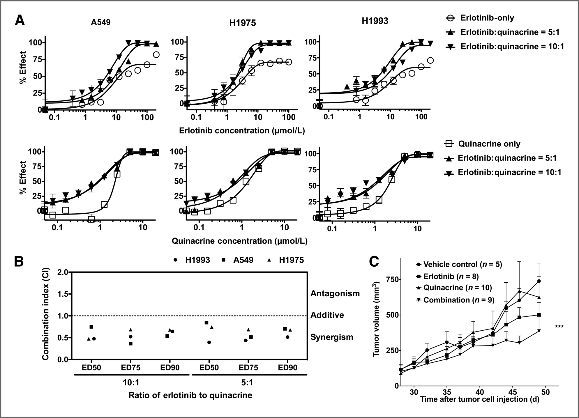
Tumorigenicity assay acrine in each cell line, which are between 5 mmol/L and NCr nu/nu athymic nude mice were obtained from 12 mmol/L and 1 to 2 mmol/L, respectively. On the basis of Taconic. Studies were conducted under an approved their IC50 ratios, we combined erlotinib with quinacrine Institutional Animal Care and Use Committee protocol at a 5 to 1 or 10 to 1 ratio and measured cell viability by the Case Comprehensive Cancer Center Athymic Ani- after treatment. We then quantified the degrees of syn- mal and Xenograft Core. A549 cells were suspended at a ergism using the median-drug effect analysis method density of 2  106 cells in 100 mL DMEM medium contain- developed by Chou and Talalay (18, 19). This method ing 5% FBS. Cell suspensions were subcutaneously quantifies the CIs of two drugs based on the growth injected into the rear flanks bilaterally of 6-week-old male inhibition curves of each drug alone or their combina- mice (n ¼ 5, 10 tumors per group). Tumor volume (mm3) tion (Fig. 1A). The combination of erlotinib and quin- was calculated with the formula 0.525  W2  L, where W acrine was synergistic in A549, H1975, and H1993 cells and L were the smallest and largest diameters of the tumor when combined at both 5:1 or 10:1 ratios [effective dose in mm, measured every other day. Tumors were grown to (ED)50: 0.61 (0.42–0.81); ED75: 0.53 (0.40–0.67); ED90: at least 200 mm3 before start of treatment. Tumors that 0.63 (0.54–0.71); Fig. 1B].
failed to engraft (reach double digit diameter) were To determine the in vivo effect of this drug combi- excluded from the study. Thereafter, mice received daily nation, we measured tumor growth in an A549 xeno- oral gavage of vehicle control (0.5% w/v methyl cellu- graft model treated with oral gavage of vehicle (0.5% lose), erlotinib (30 mg/kg/d), quinacrine (100 mg/kg methyl cellulose), erlotinib (30 mg/kg/d), and quinacrine loading dose at day one followed by 50 mg/kg/d), (50 mg/kg/d with a 2-fold initial loading dose), or a or combination of erlotinib (30 mg/kg/d) plus quina- combination of erlotinib (30 mg/kg/d) plus quinacrine crine (100 mg/kg initial dose followed by 50 mg/kg/d).
(50 mg/kg/d with a 2-fold initial loading dose). The Mice were sacrificed when tumors reached 17 mm in combination significantly inhibited in vivo tumor growth compared with vehicle control or single drug adminis-tration of quinacrine (Fig. 1C).
Statistical analysis All statistical analyses (except microarray data) were The combination of quinacrine with erlotinib induced conducted using GraphPad Prism 5. Results are repre- apoptosis and cell-cycle arrest sented by mean  SD. Statistical significance was assumed We treated A549 or H1975 cells with 1 mmol/L for a two-tailed P value less than 0.05 using ANOVA with erlotinib and either 5 or 3 mmol/L quinacrine in most the Bonferroni or Dunnett posthoc test, compared with of the subsequent experiments. The sub-IC50 concen- untreated controls or nontargeted shRNA.
tration of erlotinib was chosen because at the standarddosage of erlotinib (150 mg/d) used in the clinical setting, the maximum concentration of erlotinib (Cmax) Microarray data in the form of raw CEL and RMA achievable in humans is much lower than the IC50 normalized matrix files were deposited on the NCBI Gene of erlotinib in these resistant cell lines (26, 27). On the Expression Omnibus database under the accession num- other hand, the Cmax of quinacrine reaches 3 to 5 mmol/L ber GSE57422.
in patients (unpublished data), and quinacrine isknown to accumulate at high concentrations in tissues (especially in liver and lung) with a volume of distri- The combination of erlotinib and quinacrine is bution of approximately 50,000 L (7), and thus these synergistic in several NSCLC lung adenocarcinoma concentrations were chosen because they could be cell lines and inhibits in vivo NSCLC tumor cell achieved in vivo.
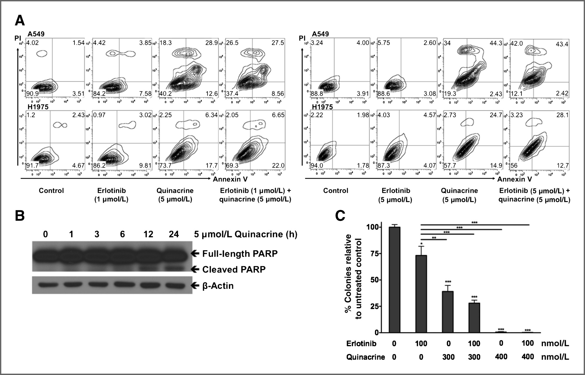
Because the combination shows synergy when erlotinib Constitutive NF-kB activation is known to mediate and quinacrine are used at their IC50 ratios (erlotinib: survival and drug resistance in cancer, and its inhibition quinacrine ¼ 5:1 or 10:1), at a concentration of 1 mmol/L has been reported to increase sensitivity to cancer thera- erlotinib and a concentration of 3 to 5 mmol/L quinacrine pies including EGFR-TKIs (12, 14). To test whether inhi- in our erlotinib-resistant cell lines, the two drugs were no bition of NF-kB is synergistic with erlotinib, a major longer synergistic and quinacrine showed potent single- EGFR-TKI used in NSCLC treatment, we tested the effects agent activity in this combination. Quinacrine alone or of the combination of erlotinib and quinacrine, an NF-kB addition of quinacrine to erlotinib induced similar levels inhibitor, on cell viability in three NSCLC cell lines: A549 of cell death, as demonstrated by increased Annexin V-PI (wtEGFR, mutant KRAS), H1975 (EGFRL858R/T790M), and staining in A549 and H1975 cells after 48 hours of treat- H1993 (MET amplification). Each of these cell lines harbor ment (Fig. 2A), and when we increased erlotinib to genetic aberrations that represent three major mechan- 5 mmol/L (which is still below its IC50), combination isms driving resistance to anti-EGFR therapy in advanced treatment induced higher levels of cell death than quin- acrine alone (Fig. 2A). We further confirmed this result by We first determined the individual IC50 values (half observing a time-dependent increase in PARP cleavage maximal inhibitory concentrations) for erlotinib and quin- induced by quinacrine (Fig. 2B). Quinacrine alone or Mol Cancer Ther; 13(9) September 2014 Molecular Cancer Therapeutics


Published OnlineFirst July 15, 2014; DOI: 10.1158/1535-7163.MCT-14-0013 Combination of Quinacrine and Erlotinib in NSCLC Figure 1. The combination of erlotinib and quinacrine is synergistic in several NSCLC lung adenocarcinoma cell lines and inhibits in vivo xenograft tumorgrowth. A, A549 (wtEGFR), H1975 (EGFR-L858R/T790M), and H1993 (MET amplification) cells were treated with erlotinib, quinacrine, or the combination ofboth agents at a 5:1 or 10:1 molar ratio. After 72 hours, cell viability was determined by the MTT assay (effect% ¼ 100%  cell viability%). The experimentswere repeated three times. B, the CI was assessed using CalcuSyn software to determine drug interaction (additivity, synergism). A CI < 1.0 isconsidered to be synergistic. All values represent mean  SD. C, growth curves of A549 lung adenocarcinoma xenograft in NCr nu/nu athymic mice. Followingan initial growth period of 35 days, group tumor volume reached at least 200 mm3 before treatment (P ¼ 0.5273 between groups). Tumor diameterswere measured every other day. Treatment continued for at least 20 days before the mice were sacrificed; , significance of P < 0.001 compared with vehiclecontrol or quinacrine only treatment (ANOVA followed by Dunnett multiple comparison test).
addition of quinacrine to erlotinib treatment also signif- phosphorylation in cells treated with either erlotinib or icantly inhibited in vitro cell proliferation, as measured by quinacrine, finding that only erlotinib inhibited AKT or colony formation (Fig. 2C).
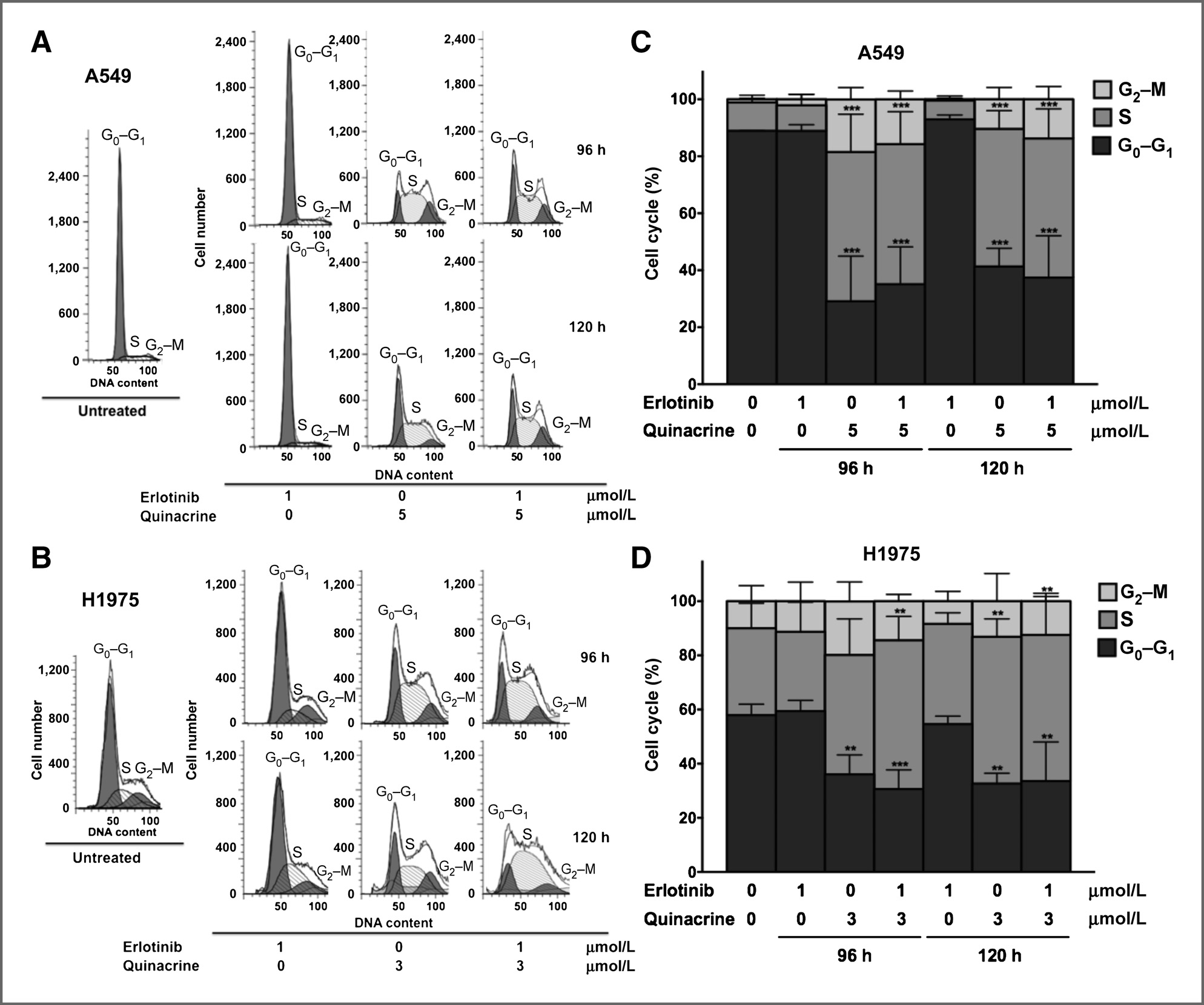
ERK activation (data not shown). This result suggests that Next, we measured the effect of quinacrine plus erlo- the effect of quinacrine on cell survival is mediated tinib on cell-cycle progression, using flow cytometry. Both through a pathway other than the PI3K/AKT or MAPK quinacrine alone and combination treatment induced similar levels of marked G1–S and G2–M cell-cycle arrest A recent report suggested that chloroquine can over- in A549 and H1975 cells. This effect was dominated come erlotinib resistance in NSCLC cells overexpressing by the action of quinacrine when the low concentration wtEGFR by inhibiting autophagy (29). Both chloroquine of 1 mmol/L erlotinib was used (Fig. 3A–D).
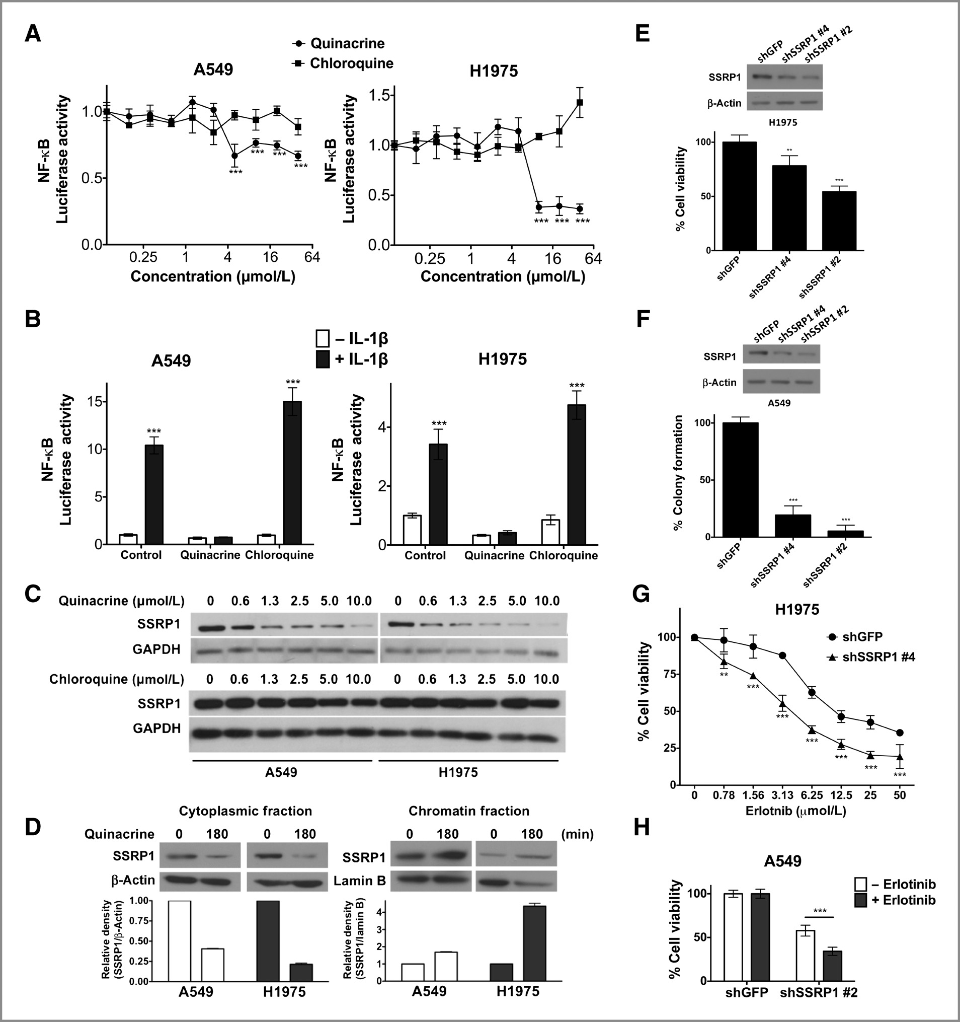
and quinacrine are known to inhibit autophagy (30), butwhether both of these antimalarial drugs inhibit NF-kB Quinacrine, but not chloroquine, suppresses NF-kB– remains uncertain. To address this issue, we determined driven luciferase activity whether quinacrine or chloroquine inhibits NF-kB activity Next, we analyzed how quinacrine overcomes erlotinib in A549 or H1975, utilizing an NF-kB-luciferase reporter resistance in NSCLC cells. Because the PI3K/AKT and assay. Luciferase expression driven by either constitutive- MAPK pathways are known to be important for cell ly active NF-kB (Fig. 4A) or IL1 treatment (Fig. 4B) was survival and are determinants of EGFR-TKI sensitivity significantly suppressed by quinacrine but not by equal in EGFR-driven cancers (28), we analyzed AKT and ERK concentrations of chloroquine.
Mol Cancer Ther; 13(9) September 2014


Published OnlineFirst July 15, 2014; DOI: 10.1158/1535-7163.MCT-14-0013 Dermawan et al.
Figure 2. The combination of quinacrine with erlotinib induced in vitro NSCLC cell apoptosis and inhibited cell growth. A, A549 and H1975 cells were untreatedor treated with 1 mmol/L (left) or 5 mmol/L (right) erlotinib, 5 mmol/L quinacrine, or both for 48 hours. Apoptosis was assessed by Annexin V-PI double stainingfollowed by flow-cytometric analysis. B, H1975 cells were treated with 5 mmol/L quinacrine for the indicated time-points. Western analysis was used todetect PARP cleavage as an indicator of apoptosis. C, A549 cells were seeded in 6-well plates and treated with erlotinib, quinacrine, or a combination of both intriplicate at the indicated concentrations. Drugs were replaced every 72 hours. After 14 days, colonies were stained and quantified. Statistical analysis of thedifferences in colony formation between the treated cells and untreated controls were conducted using one-way ANOVA followed by Bonferroni multiplecomparison test (, P < 0.05; , P < 0.01; , P < 0.001). All values represent mean  SD.
Quinacrine mediates cell killing and overcomes as shown by the ability of quinacrine, but not chloroquine, resistance to erlotinib by targeting FACT to reduce mobility of plasmid DNA (Supplementary Gasparian and colleagues showed that a series of anti- Fig. S1C). To test whether the anticancer activity of quin- cancer compounds including quinacrine suppresses NF- acrine in NSCLC is due to inhibition of FACT, we knocked kB activation by causing chromatin trapping of the FACT SSRP1 down in A549 or H1975 cells. Loss of SSRP1 complex (10). This finding is supported by our observa- significantly decreased cell survival (Fig. 4E and F) and tion that treatment of A549 or H1975 cells with quinacrine, increased sensitivity to erlotinib (Fig. 4G and H) in both but not chloroquine, rapidly depletes SSRP1, a FACT subunit, from the soluble cytoplasmic fraction (Fig. 4Cand D), and led to SSRP1 accumulation in the insoluble The quinacrine and erlotinib combination inhibits chromatin fraction (Fig. 4D), which has also been shown the expression of SSRP1-regulated genes and cell- by Gasparian and colleagues to be an indicator of chro- cycle genes that predict worse survival in patients matin trapping of FACT (10). Because the overall level of with lung adenocarcinoma SSRP1 from whole-cell lysates remained unchanged (Sup- To further elucidate the mechanisms of the effect of plementary Fig. S1A), the decrease of SSRP1 from the combined quinacrine and erlotinib treatment in NSCLC, cytoplasmic fraction was not due to protein degradation.
we performed global transcriptomic profiling of A549 and Quinacrine and chloroquine are structurally related com- H1975 cells treated with 1 mmol/L erlotinib alone, 5 or pounds known to interact with DNA, but with different 3 mmol/L quinacrine alone, or combinations of 1 mmol/L affinities, due to the stronger drug-DNA ring–ring stack- erlotinib and 5 or 3 mmol/L quinacrine for 0, 6, 12, ing interaction with quinacrine, which has a 3-ring acri- or 24 hours, using an Affymetrix microarray platform.
dine moiety, compared with chloroquine, which has a 2- (48-hour treatment samples were available for erlotinib ring quinolone moiety (Supplementary Fig. S1B; ref. 31), alone or combination treatment). Principal component Mol Cancer Ther; 13(9) September 2014 Molecular Cancer Therapeutics


Published OnlineFirst July 15, 2014; DOI: 10.1158/1535-7163.MCT-14-0013 Combination of Quinacrine and Erlotinib in NSCLC Figure 3. The combination of quinacrine and erlotinib inhibits G1–S and G2–M cell-cycle progression. A549 (A and C) and H1975 (B and D) cells were untreatedor treated with erlotinib only, quinacrine only, or erlotinib plus quinacrine for 72 or 96 hours at the indicated concentrations. Cell-cycle analysis was thenperformed using PI-staining followed by flow-cytometric analysis. G1–S and G2–M cell-cycle arrest was determined by quantifying relative G0–G1, S,and G2–M phase percentages. Statistical analysis of the differences in relative cell-cycle phase percentages between the treated cells and untreated controlswere conducted using two-way ANOVA followed by Dunnett multiple comparison test (, P < 0.01; , P < 0.001). The experiment was repeated three times.
All values represent mean  SD.
analysis revealed that, relative to untreated cells at 0 hour, ing our functional analysis showing that quinacrine plus gene expression profiles diverged most significantly with erlotinib induced significant cell-cycle arrest and inhib- increased treatment time (24 and 48 hours) and with ited tumor growth.
quinacrine or combination treatment (Supplementary Next, we analyzed the enrichment of TFBSs among Fig. S2). We next determined genes that were differen- those genes whose expression was significantly affected tially expressed between treatment groups. Differential by quinacrine or by erlotinib plus quinacrine. Comparison gene expression analysis was used to identify genes that of our data with the ChIP-SEQ results for SSRP1-enriched were significantly induced or suppressed by combination genes reported by Garcia and colleagues (16) showed that treatment compared with either single drug treatment many of the genes affected by quinacrine or combination and whose expression levels showed a >2-fold change treatment were regulated by the same transcription fac- relative to 0 hour and significant temporal profiles (Fig. 5A tors that were also involved in regulating expression of and B). Gene ontology analysis showed that genes signif- SSRP1-enriched genes. These transcription factors belong icantly affected by the combination were most highly to the EGR (EGR1), ETS (ELK1, ELK4, GABPA, SPI1), enriched for those encoding proteins involved in cell- MYC (MYC, MYCN), and SP/KLF (SP1, KLF4) families cycle progression or DNA metabolism (Table 1), confirm- (Supplementary Table S1). This result supports our Mol Cancer Ther; 13(9) September 2014


Published OnlineFirst July 15, 2014; DOI: 10.1158/1535-7163.MCT-14-0013 Dermawan et al.
Figure 4. Quinacrine, but not chloroquine, suppresses NF-kB–driven luciferase activity, mediates cell killing, and overcomes resistance to erlotinib by targetingFACT. A, luciferase units relative to untreated control (RLU) were quantified in A549 or H1975 cells stably expressing NF-kB luciferase reporter after 4 hours oftreatment with increasing concentrations of quinacrine or chloroquine in triplicate. Statistical analysis of the differences in RLU between cells treated withdifferent drug concentrations and untreated controls were conducted using two-way ANOVA followed by Dunnett multiple comparison test (, P < 0.001).
B, RLU was quantified in A549 or H1975 stable NF-kB luciferase reporter cells pretreated with 10 mmol/L quinacrine or 10 mmol/L chloroquine for 1hour, andthen stimulated with 10 ng/mL IL1b for 6 hours in quadruplicate. Statistical analysis of the differences in RLU between IL1-treated or -untreated cellswere conducted using two-way ANOVA followed by Bonferroni posttest (, P < 0.001). All values represent mean  SD. C, A549 or H1975 cells weretreated with quinacrine or chloroquine for 1 hour at the indicated concentrations. Soluble protein fractions were then extracted by mild cell lysis and theSSRP1 subunit of the FACT complex was analyzed by the Western method. D, A549 or H1975 cells were treated with 20 mmol/L quinacrine for 3 hours. Thelevels of SSRP1 in the cytoplasmic and chromatin fractions were analyzed by the Western method and quantified by densitometry. b-actin or lamin B served asloading controls. E, H1975 cells were transduced with shRNA lentiviruses against GFP or SSRP1. Cells were plated in quadruplicate and cell viabilitywas measured by MTT assay 5 days after infection. F, A549 cells were infected with shRNA against GFP or SSRP1. Cells were plated in 6-well plates intriplicate and cell colonies were quantified after 2 weeks by crystal violet staining. H1975 (G) or A549 (H) cells were transiently infected with shSSRP1 andplated in 96-well plates and treated with DMSO or increasing concentrations of erlotinib over 72 hours in quadruplicate. Cell viability was measured by MTTassay. Statistical analyses of the differences in cell viability or colony formation between SSRP1 or GFP knockdown cells were conducted using one-way ortwo-way ANOVA followed by Dunnett multiple comparison test (, P < 0.01; , P < 0.001). All values represent mean  SD.
Mol Cancer Ther; 13(9) September 2014 Molecular Cancer Therapeutics Published OnlineFirst July 15, 2014; DOI: 10.1158/1535-7163.MCT-14-0013 Combination of Quinacrine and Erlotinib in NSCLC Figure 5. Quinacrine and erlotinib combination treatment inhibits SSRP1-regulated genes and cell-cycle genes whose increased expression predicts poorersurvival in patients with lung adenocarcinoma. A549 (top) or H1975 (bottom) cells were treated with or without erlotinib, quinacrine, or combination for 6, 12, or24 hours. Differential gene expression analysis was used to identify genes that were significantly induced or suppressed by combination treatment comparedwith single drug treatment and showed a >2-fold change relative to 0 hour and significant temporal profiles, as represented by Venn diagrams (A) andhierarchical clustering (B). C, as potential biomarkers of drug combination efficacy in the ongoing clinical trial, cell-cycle–related genes that showed significanttemporal suppression by combination treatment compared with either single treatment alone in A549 cells were identified and represented in a heatmap. D,examples of the Kaplan–Meier survival curves of patients with lung adenocarcinoma from Supplementary Table S2 with high versus low KIFC1, FOSL1, BIRC5, or HIST1H2BM expression.
observation that quinacrine targets and inhibits the FACT in patients with NSCLC (HR ranges from 1.19 to 1.98), complex (16). Interestingly, the levels of the FACT subunit and this correlation was even more significant when SSRP1 and SPT16 mRNAs were not affected by drug only patients with lung adenocarcinoma were analyzed treatment in our microarray study (data not shown), (HR ranges from 1.58 to 2.92; Fig. 5D and Supplemen- which corroborates previous reports showing that the tary Table S2). This result is relevant to an ongoing action of quinacrine on FACT is at a functional level, by phase I/II clinical trial (NCT01839955) to test the com- trapping the FACT complex onto chromatin.
bination of erlotinib and quinacrine in metastatic (stage To identify potential biomarkers for erlotinib-quina- IIIB-IV) NSCLC patients who failed first-line chemo- crine synergy, we identified genes that were suppressed therapy, the vast majority of whom have wtEGFR non– more significantly by combination treatment than by small cell lung adenocarcinoma tumors. Therefore, our either drug alone in A549 lung adenocarcinoma cells preclinical studies for this clinical trial not only identify (Fig. 5C). The more potent suppression of this gene set a potential set of treatment response pharmacodynamic by combination treatment was verified by TaqMan- biomarkers, but also suggest important biologic based qRT-PCR analysis (Supplementary Fig. S3).
mechanisms regulating the potent single-agent activity Because our functional analysis showed that the com- of quinacrine activity in erlotinib-insensitive NSCLC bination of erlotinib and quinacrine induced significant cell-cycle arrest, we preferentially selected genes thatour gene ontology analysis showed to be involved in cell-cycle progression. Importantly, increased expres- Erlotinib is effective in patients with NSCLC with sion of these genes was associated with poorer survival known drug-sensitizing EGFR mutations (3), but its Mol Cancer Ther; 13(9) September 2014 Published OnlineFirst July 15, 2014; DOI: 10.1158/1535-7163.MCT-14-0013 Dermawan et al.
Table 1. Gene ontology processes for genes that are significantly affected by erlotinib-quinacrinecombination treatment Cell-cycle process M phase of mitotic cell cycle Mitotic cell cycle Organelle fission DNA metabolic process Response to DNA damage stimulus Cellular response to stress Chromosome segregation Negative regulation of macromolecule biosynthetic process Notch signaling pathway Negative regulation of macromolecule metabolic process Negative regulation of transcription Regulation of RNA metabolic process Pyrimidine deoxyribonucleotide metabolic process Negative regulation of gene expression Regulation of DNA metabolic process clinical efficacy in patients with wtEGFR or acquired and treatment of malaria and other parasitic diseases, are resistance to TKIs due to secondary mutations remains well tolerated (7, 8). A future experiment looking at modest (3, 6). We show here that the addition of quina- whether overexpression of FACT itself could overcome crine to erlotinib in several patient-derived erlotinib-resis- resistance to erlotinib or other EGFR-TKI in NSCLC tant NSCLC cell lines overcomes resistance to erlotinib. A would be valuable.
major advantage of our strategy is the pairing of a highly We are currently conducting a phase I/II clinical trial specific small-molecule kinase inhibitor, erlotinib, to a (NCT01839955) to test erlotinib alone versus erlotinib plus broadly acting DNA intercalator, quinacrine, thereby quinacrine in patients with locally advanced or metastatic decreasing the chance of emergence of resistance against (stage IIIB/IV) NSCLC with either: (i) wtEGFR, with targeted therapies.
disease progression after previous platinum-based che- Quinacrine has been shown to reduce the availability of motherapy, for which erlotinib was approved as a second- the FACT complex by causing its trapping on chromatin line monotherapy; or (ii) documented EGFRL858R/T790M (10), and FACT was shown by the same group to promote mutation or EML4–ALK fusion gene, with subsequent tumor survival and growth (16). Because the ability of progression on first-line erlotinib or crizotinib and che- quinacrine to inhibit FACT and subsequently modulate motherapy. Using the Chou–Talalay method, we deter- NF-kB–dependent transcriptional activity is not depen- mined the two drugs to be synergistic at their respective dent on direct binding to these targets but is mediated by IC50 ratios in erlotinib-resistant cell lines, when erlotinib binding to DNA (10), it may be less likely for drug was used at a much higher concentration (5:1 or 10:1) than resistance to arise. Even though quinacrine intercalates quinacrine. However, based on preexisting pharmacoki- into DNA, it is not genotoxic (10), and its side effects and netics data, in these erlotinib-resistant patients, erlotinib is general toxicity, which have been well documented over likely to reach only a sub-IC50 serum concentration (26, several decades due to its extensive use in the prevention 27), whereas quinacrine is known to accumulate in tissues Mol Cancer Ther; 13(9) September 2014 Molecular Cancer Therapeutics Published OnlineFirst July 15, 2014; DOI: 10.1158/1535-7163.MCT-14-0013 Combination of Quinacrine and Erlotinib in NSCLC (7). When we selected drug doses more representative of independent pathways is exponentially smaller than the those achievable clinically, we observed potent single- probability of resistance to a single agent. Our novel agent activity of quinacrine in the erlotinib-resistant combination offers a promising therapy in advanced NSCLC cell lines. At such concentrations, quinacrine NSCLC, for which there are currently few effective treat- dominates the cell killing activity of the combination, as ment options after the tumors have progressed during indicated by our colony formation, cell cycle, and apo- first-line anticancer treatments.
ptosis assay, and our drug synergy quantification predictsthat a much higher erlotinib concentration would be Disclosure of Potential Conflicts of Interest needed to achieve synergy with quinacrine in these N. Sharma received a commercial research grant from Incuron Inc. No assays. This result suggests that the inclusion of quina- potential conflicts of interest were disclosed by the other authors.
crine alone as one of the arms in a future clinical trialwould be valuable. Our findings also indicate that obser- Authors' ContributionsConception and design: J.K.T. Dermawan, K. Gurova, A. Dowlati, S. De, vation of drug synergy in in vitro studies might fail to G. Narla, N. Sharma, G. Stark translate to clinical trials when the same ratios are not Development of methodology: J.K.T. Dermawan, K. Gurova, A. Dowlati, attainable based on in vivo drug pharmacokinetics.
S. De, G. Narla, G. StarkAcquisition of data (provided animals, acquired and managed patients, We also identified, as potential pharmacodynamic mar- provided facilities, etc.): J.K.T. Dermawan, A. Dowlati, N. Sharma kers for this clinical trial, a set of genes whose expression Analysis and interpretation of data (e.g., statistical analysis, biostatis- levels were significantly suppressed by combination ther- tics, computational analysis): J.K.T. Dermawan, J.J. Pink, A. Dowlati,G. Narla, N. Sharma, G. Stark apy and were shown to correlate with worse patient Writing, review, and/or revision of the manuscript: J.K.T. Dermawan, survival in existing gene expression databases. Because A. Dowlati, S. De, G. Narla, N. Sharma, G. StarkAdministrative, technical, or material support (i.e., reporting or orga- these are posttreatment rather than pretreatment biomar- nizing data, constructing databases): J.J. Pink, N. Sharma, G. Stark kers, the goal is to use these gene signatures to validate Study supervision: N. Sharma, G. Stark that the biologic efficacy of the combination over singledrug treatment during the early phase of treatment by gene expression profiling of pre- and posttreatment biop- The authors thank Drs. Martina Veigl and Patrick Leahy at the Gene Expression Array Core Facility for their valuable input in the design and sies from the subjects in our clinical trial, and then apply analysis of the microarray study, Vai Pathak for conducting the ABI this knowledge to select for patients who would truly Expression Real-time RT-PCR experiments, Ian Lent at the Translational benefit from the combination and thus would remain in Research & Pharmacology Core Facility for performing the CalcuSynanalysis, and Cathy Shemo and Bunny Cotleur at the CCF Flow Cytometry the trial through the entire course of treatment. For future Core for excellent technical support for flow-cytometric data acquisition studies, to identify pretreatment predictive pharmacody- and analysis.
namics biomarkers, we would need to identify cell linesthat are resistant to the combination and compare their gene signatures with those of cell lines that are sensitive to This work was supported by the National Cancer Institute Grant P01 CA062220 (to G.R. Stark), Clinical & Translational Science Collaborative of the combination, such as those used in the present study.
Cleveland Core Utilization pilot grant P30 CA043703-23 at Case Western Recent discoveries of the high degree of intratumoral Reserve University (to N. Sharma), and a Harrington Discovery Institute and intermetastatic genetic heterogeneity among tumor grant (to G. Narla).
The costs of publication of this article were defrayed in part by the cells in cancer genomics projects suggest that the devel- payment of page charges. This article must therefore be hereby marked opment of resistance is inevitable in any targeted therapy advertisement in accordance with 18 U.S.C. Section 1734 solely to indicatethis fact.
for cancer (32). The use of combinatorial therapy is animportant means to circumvent this problem because the Received January 10, 2014; revised May 29, 2014; accepted June 23, 2014; probability of cancer cells becoming resistant to two published OnlineFirst July 15, 2014.
Lynch TJ, Bell DW, Sordella R, Gurubhagavatula S, Okimoto RA, lung tumors with acquired resistance to gefitinib or erlotinib. Proc Natl Brannigan BW, et al. Activating mutations in the epidermal growth Acad Sci U S A 2007;104:20932–7.
factor receptor underlying responsiveness of non-small cell lung Sequist LV, Waltman BA, Dias-Santagata D, Digumarthy S, Turke AB, cancer to gefitinib. N Engl J Med 2004;350:2129–39.
Fidias P, et al. Genotypic and histological evolution of lung cancers Garassino MC, Martelli O, Broggini M, Farina G, Veronese S, Rulli E, acquiring resistance to EGFR inhibitors. Sci Transl Med 2011;3: et al. Erlotinib versus docetaxel as second-line treatment of patients with advanced non-small cell lung cancer and wild-type EGFR tumors Ehsanian R, Van Waes C, Feller SM. Beyond DNA binding – a review of (TAILOR): a randomized controlled trial. Lancet Oncol 2013;14:981–8.
the potential mechanisms mediating quinacrine's therapeutic activi- Shepherd FA, Rodrigues PJ, Ciuleanu T, Tan EH, Hirsh V, Thongpra- ties in parasitic infections, inflammation, and cancers. Cell Commun sert S, et al. Erlotinib in previously treated non-small cell lung cancer. N Signal 2011;9:13.
Engl J Med 2005;353:123–32.
Gurova K. New hopes from old drugs: revisiting DNA-binding small Kobayashi S, Boggon TJ, Dayaram T, Janne PA, Kocher O, Meyerson molecules as anticancer agents. Future Oncol 2009;5:1685–704.
M, et al. EGFR mutation and resistance of non-small cell lung cancer to Gurova KV, Hill JE, Guo C, Prokvolit A, Burdelya LG, Samoylova E, et al.
gefitinib. N Engl J Med 2005;352:786–92.
Small molecules that reactivate p53 in renal cell carcinoma reveal a NF- Bean J, Brennan C, Shih JY, Riely G, Viale A, Wang L, et al. MET kB-dependent mechanism of p53 suppression in tumors. Proc Natl amplification occurs with or without T790M mutations in EGFR mutant Acad Sci U S A 2005;102:17448–53.
Mol Cancer Ther; 13(9) September 2014 Published OnlineFirst July 15, 2014; DOI: 10.1158/1535-7163.MCT-14-0013 Dermawan et al.
10. Gasparian AV, Burkhart CA, Purmal AA, Brodsky L, Pal M, Saranadasa 22. Ernst J, Bar-Joseph Z. STEM: a tool for analysis of short time series M, et al. Curaxins: anticancer compounds that simultaneously sup- gene expression data. BMC Bioinformatics 2006;7:191.
press NF-kB and activate p53 by targeting FACT. Sci Transl Med 23. Sherman BT, Huang DW, Lempicki RA. Systemic and integrative analysis of large gene lists using DAVID bioinformatics resources. Nat 11. Lu T, Sathe SS, Swiatkowski SM, Hampole CV, Stark GR. Secretion of cytokines and growth factors as a general cause of constitutive NFkB 24. Ho Sui SJ, Mortimer JR, Arenillas DJ, Brumm J, Walsh CJ, Kennedy activation in cancer. Oncogene 2004;23;2138–45.
BP, et al. oPOSSUM: identification of over-represented transcription 12. Nakanishi C, Toi M. Nuclear factor-kappaB inhibitors as sensitizers to factor binding sites in co-expressed genes. Nucleic Acid Res anticancer drugs. Nat Rev Cancer 2005;5:297–309.
13. Tanaka K, Babic I, Nathanson D, Akhavan D, Guo D, Gini B, et al.
 rffy B, Surowiak P, Budczies J, Lanczky A. Online survival analysis Oncogenic EGFR signaling activates an mTORC2-NF-kB pathway that software to assess the prognostic value of biomarkers using tran- promotes chemotherapy resistance. Cancer Discov 2011;1:524–38.
scriptomic data in non-small-cell lung cancer. PLoS ONE 2013;8: 14. Bivona TG, Hieronymus H, Parker J, Chang K, Taron M, Rosell R, et al.
FAS and NF-kB signaling modulate dependence of lung cancers on 26. Hidalgo M, Siu LL, Neumunaitis J, Rizzo J, Hammond LA, Takimoto C, mutant EGFR. Nature 2011;471:523–6.
et al. Phase I and pharmacologic study of OSI-774, an epidermal 15. Brown M, Cohen J, Arun P, Chen Z, Van Waes C. NF-kB in carcinoma growth factor receptor tyrosine kinase inhibitor, in patients with therapy and prevention. Exp Opin Ther Targets 2008;12:1109–22.
advanced solid malignancies. J Clin Oncol 2001;19:3267–79.
16. Garcia H, Miecznikowski JC, Safina A, Commane M, Ruusulehto A, 27. Lu JF, Eppler SM, Wolf J, Hamilton M, Rakhit A, Bruno R, et al. Clinical Kilpinen S, et al. Facilitates Chromatin Transcription Complex is an pharmacokinetics of erlotinib in patients with solid tumors and expo- "accelerator" of tumor transformation and potential marker and target sure-safety relationship in patients with non-small cell lung cancer.
of aggressive cancers. Cell Rep 2013;4:159–73.
Clin Pharmacol Ther 2006;80:136–45.
17. van Meerloo J, Kaspers GJ, Cloos J. Cell sensitivity assays: the MTT 28. Sharma SV, Bell DW, Settleman J, Haber DA. Epidermal growth factor assay. Methods Mol Biol 2011;731:237–45.
receptor mutations in lung cancer. Nat Rev Cancer 2007;7:169–81.
18. Chou TC. Drug combination studies and their synergy quantification 29. Zou Y, Ling YH, Sironi J, Schwartz EL, Perez-Soler R, Piperdi B. The using the Chou-Talalay method. Cancer Res 2010;70:440–6.
autophagy inhibitor chloroquine overcomes the innate resistance of 19. Chou TC, Talalay P. Quantitative analysis of dose-effect relationships: wild-type EGFR non-small-cell-lung cancer cells to erlotinib. J Thorac the combined effects of multiple drugs or enzyme inhibitors. Adv Enzyme Regul 1984;22:27–55.
30. Kimmelman AC. The dynamic nature of autophagy in cancer. Genes 20. Gasparian AV, Guryanova OA, Chebotaev DV, Shishkin AA, Yemelya- nov AY, Budunova IV. Targeting transcription factor NF-kB: compar- 31. Bolte J, Demuynck C, Lhomme MF, Lhomme J, Barbet J, Roques BP.
ative analysis of proteasome and IKK inhibitors. Cell Cycle 2009;8: Synthetic models related to DNA intercalating molecules: comparison between quinacrine and chloroquine in their ring-ring interaction with 21. Ishwaran H, Rao JS, Kogalur UB. BAMarray: Java software for Bayes- adenine and thymine. J Am Chem Soc 1982;104:760–65.
ian analysis of variance for microarray data. BMC Bioinformatics 32. Garraway LA, Lander ES. Lessons from the cancer genome. Cell Mol Cancer Ther; 13(9) September 2014 Molecular Cancer Therapeutics Published OnlineFirst July 15, 2014; DOI: 10.1158/1535-7163.MCT-14-0013 Quinacrine Overcomes Resistance to Erlotinib by Inhibiting FACT,
NF-

Small Cell Lung Cancer
B, and Cell-Cycle Progression in Non
Josephine Kam Tai Dermawan, Katerina Gurova, John Pink, et al. 2014;13:2203-2214. Published OnlineFirst July 15, 2014.
Mol Cancer Ther Access the most recent version of this article at: Access the most recent supplemental material at: This article cites 32 articles, 8 of which you can access for free at: This article has been cited by 3 HighWire-hosted articles. Access the articles at: Reprints and
To order reprints of this article or to subscribe to the journal, contact the AACR Publications Department at To request permission to re-use all or part of this article, contact the AACR Publications Department at

Source: http://incuron.ru/doc/st/13.pdf

Isc310343.qxd

and Developing Academic and Behavioral Interventions for Students with Bipolar KIM KILLU AND R. MARK A. CRUNDWELL Despite significant advances in practices for effectively designing anddelivering instruction for students with disabilities, educators continueto face challenges addressing the needs of students with emotionaland behavioral disorders. Little information is available for educatorson accommodations and modifications that would serve the needs ofthese students and address the unique challenges they present in theclassroom. The educational, social, and behavioral needs of studentswith bipolar disorder are discussed along with suggestions for provid-ing effective accommodations and modifications in the classroom.

Microsoft word - liberty global.doc

Liberty Global, Inc. Programa de conducta empresarial y cumplimiento del reglamento Este documento proporciona información sobre nuestro Código de conducta empresarial. Todos los directores, gerentes y empleados son responsables, individual y colectivamente, de la gestión de nuestras actividades comerciales conforme al Código y a la legislación pertinente. Por ello, le recomendamos que lea este documento con atención. Al aceptar un cargo o continuar ejerciendo sus funciones como director, gerente o empleado, se compromete a cumplir lo establecido en el presente Código. El desconocimiento del Código no exime a ningún empleado de la responsabilidad de atenerse a él, así como a la legislación pertinente y a cualquier otra normativa aplicable a sus actividades laborales.