Even if Viagra is not needed, it is possible that the doctor will be able to determine the etiology of erectile dysfunction and prescribe appropriate treatmen https://vgraustralia.net it doesn't pay to forget about sexual activeness even at the first sings of malfunction.
Gwk-munich.de
Microchim ActaDOI 10.1007/s00604-011-0548-9
Simultaneous determination of four different antibioticresidues in honey by chemiluminescence multianalytechip immunoassays
Klaus Wutz & Reinhard Niessner & Michael Seidel
Received: 9 November 2010 / Accepted: 14 January 2011
# Springer-Verlag 2011
Abstract We are presenting the first method for identifi-
Keywords Antibiotic microarray . Chemiluminescence
cation and quantification of antibiotic derivatives in honey
detection . Regenerable biochip . Automated flow-injection
samples using regenerable antigen microarrays in combi-
system . Microarray image evaluation
nation with an automated flow injection system. Thescheme is based on an indirect competitive immunoassayformat using monoclonal antibodies bound to the surface of
the microarray. The surface of glass slides was coated withepoxy-activated poly(ethylene glycol) and enables direct
Honey is generally considered as natural and healthy
immobilization of the antibiotic derivatives. The antigen/
product of animal origin. However, in the recent years
antibody interaction on the surface of the chip can be
there were some publications dealing with the determina-
detected by chemiluminescence (CL) read-out via CCD
tion of antimicrobial contaminants in bee products. Anti-
camera. The method allows for fast analysis of the four
biotics are used in apiculture for the treatment of bacterial
analytes simultaneously and without purification or extrac-
diseases, in particular American and European foulbrood
tion. An effective data evaluation method also was
For the effective abatement of the causers Paenibacillus
developed to warrant unambiguous identification of the
larvae and Melissoccocus pluton, respectively, drugs based
spots and to establish grey levels of CL intensities. The
on antibiotic derivatives, e.g. sulfonamides and tetracy-
software developed enables fast and automated processing
clines, have been approved All sulfonamides inhibit
of the CL images. Dose–response curves were obtained for
the bacterial synthesis of folic acid due to their structural
the derivatives of enrofloxacin, sulfadiazine, sulfametha-
analogy to p-aminobenzoic acid, whereas the group of
zine and streptomycin. Spiking experiments revealed
tetracylines interferes with the ribosomal protein synthesis.
adequate recoveries within the dynamic ranges of the
The broad activity spectrum of these antimicrobials has led
calibration curves of enrofloxacin (92%±6%), sulfametha-
to widely use in veterinary practice since the 1950s
zine (130%±21%), sulfadiazine (89%±20%) and strepto-
implicating the appearance of bacterial resistance. Hence,
mycin (93% ±4%).
new synthetic agents, e.g. the class of quinolones, havebeen developed to replace effectless antibiotics. The use ofantibiotics for the treatment of honey bees is illegal in theEuropean Union, but due to the high import quota from
Electronic supplementary material The online version of this article
other countries, contaminated honey products can be found
contains supplementary material,
on the European markets In the last years another
which is available to authorized users.
source for the contamination of honey with antibiotic
K. Wutz : R. Niessner : M. Seidel (*)
residues has been attracting notice. The plant disease
Institute of Hydrochemistry and Chair for Analytical Chemistry,
fireblight has become a serious problem for fruit-growers.
Technische Universität München,
Fireblight is caused by the bacterium Erwinia amylovora
Marchioninistraße 17,
which affects plants from the family of rosaceae, e.g. apple
81377 München, Germanye-mail:
[email protected]
and pear. This infection disease leads to dieback of the
K. Wutz et al.
blossoms and even can cause death of the entire tree [].
flow injection system []. Analytical microarrays are a
The associated great economic loss for fruit-growers
powerful tool for the simultaneous detection of multiple
requires effective abatement of Erwinia amylovora. Up to
analytes in a single measurement due to separated affinity-
now, agents based on the aminoglycoside streptomycin are
binding events at the surface interface, which reduces time
the most common treatment of affected plants and orchards
and costs for the analysis of contaminations in food [
Thus, antibiotic residues can be found in honey bee
They have been approved for the detection of DNA target
products due to contaminated collected pollen.
molecules [as well as for e.g. microorganisms ,
As contamination of food with antibiotic residues poses
and toxins [or pharmaceuticals [in food and water
risks for human health particularly with regard to increasing
samples. For analytical microarray read-out purposes
formation of resistance in bacteria strains to antimicrobials,
fluorescence []- and chemiluminescence (CL) [
there is a need for monitoring of antibiotic contamination in
detection methods are often used [, ]. CL microarrays
honey. Most of the screening methods which have been
measure the light emitted by an enzyme-assisted chemical
published are using chromatographic techniques. In partic-
reaction. In contrast to fluorescence, there is no background
ular, liquid chromatography with fluorescence detection or
signal, neither from the light source nor from light
combined with coupled mass spectrometry detection (LC-
scattering from the matrix. Therefore, CL is the most sen-
MS/MS) is used for the determination of antibiotics in food
sitive read-out principle for microarrays ,
matrices. In most cases, multi-analyte screening methods
Since microarray-immunoassay methods require several
are only applicable for antimicrobials of similar molecular
incubation and washing steps, miniaturized bioanalysis
structures. There were methods published for the determi-
platforms have been developed which combine microarray
nation of several macrolides sulfonamides [] and
assays with fluidic microsystems , ]. Using flow-
tetracyclines in honey. Some chromatographic multi-
through microarrays, the microarray is part of a flow cell
class methods have been established in the past years which
through which samples and reagents are pumped. In
allow identification [and also quantification [] of a
comparison with conventional micro-well plates, flow-
broad spectrum of antibiotics in honey. All HPLC based
through microarrays present thinner diffusion layers en-
methods require liquid-liquid or solid-phase extraction of
abling efficient mass transport [This reduces the time
the matrix followed by pre-concentration or clean-up of the
needed to perform a multianalyte assay. Thus, automated
extract. For multiclass screening there is the additional need
flow-through microarrays allow the analysis of a sample
of several extraction steps in dependency on polarity and
within minutes and ensure reproducible and easy operating.
solubility of the various analyte structures. This sample
The design of the platform MCR 3 (Microarray Chip
preparation is time-consuming as well as laborious and can
Reader 3), which is used in this study, has been published
lead to loss of analytes along the extraction procedure. The
for the determination of antibiotic residues in milk [
development of an adequate chromatographic separation of
The specific antibody/antigen interactions on the regener-
a broad variety of antibiotic residues is also a challenge
able microarray chip surface allow for determination of
which requires optimization. Additionally, in case of honey
different analytes in parallel without extraction and separa-
strong matrix effects due to the floral ingredients can cause
tion steps before analysis. The detection is carried out by
interferences which have to be minimized for chromato-
sensitive CL read-out using horseradish peroxidase (HRP)-
graphic analysis ].
Thus, chromatographic methods are not preferable for
fast and cost-efficient high-throughput monitoring.
The advantage of immunochemical methods is the usage
Materials and methods
of specific antibodies to the analyte, which is dedicated formultianalyte screening. Various studies have been
Chemicals and reagents
concerned with the immunological screening for antimicro-bial substances in honey and other food matrices [].
The water used for all aqueous buffer solutions was
Otherwise, as heterogeneous immunochemical methods are
deionized and treated by a Milli-Q plus 185 system
based on the formation of the antigen/antibody complex on
(Millipore, Schwalbach, Germany,
solid phase, time-consuming incubation steps are necessary.
All standard chemicals for the production of buffer
Because of this, methods based on ELISA formats executed
solutions were obtained from Sigma-Aldrich (Taufkirchen,
in standard micro-well plates are not applicable for fast
Germany, ). The antibiotic deriva-
screening of high sample amounts. Therefore, at our
tives streptomycin sulfate, sulfadiazine (SDZ) sodium salt,
institute we have been focusing on microarray technique
sulfamethazine (SMZ) sodium salt and enrofloxacin were
based on multianalyte immunoassays (MIA) in combination
purchased from Sigma-Aldrich. Clinafloxacin hydrochlo-
with a flow-through principle realized by construction of a
ride was obtained from Axxora (Lörrach, Germany,
Simultaneous determination of four antibiotic residues in honey
). The positive control N-(2,4-dinitrophenyl)-
slides were functionalized with 0.6 mL molten diamino-
ethylene diamine (DNPEDA) was purchased from Chem-
PEG for one sandwich in smelter at 98 °C for 15 h. The
Pur (Karlsruhe, Germany,
resulting diamino-PEG-coated glass slides were washed
For surface modification 3-glycidyloxypropyltrimethoxysilane
with water and dried under a nitrogen flow. The diamino-
(GOPTS) and poly(ethylene glycol)diglycidyl ether
PEG chips were stored at room conditions for a maximum
(diepoxy-PEG) were obtained from Sigma-Aldrich.
Diamino-poly(ethylene glycol) (diamino-PEG) was pro-
Diepoxy-PEG glass slides were prepared by dispensing
vided by Huntsman Holland (Rozenburg, The Netherlands,
0.6 mL diepoxy-PEG on one diamino-PEG glass slide and
covering it with another diamino-PEG glass slide. The
The monoclonal primary antibodies (mAb) used for the
slides were incubated in a sandwich format for 15 h at
detection of norfloxacin (mAb 1F7, reactive with enroflox-
100 °C. After cleaning with methanol and drying under
acin), streptomycin (mAb 4E2), sulfamethazine (mAb 4D9)
nitrogen the produced diepoxy-PEG-coated glass slides
and sulfadiazine (mAb 2G6) were produced at the Chair of
were directly used for the spotting process.
Hygiene and Technology of Milk (LMU München) ].
For spotting, the antibiotic derivatives were dissolved in
The mouse monoclonal antibody to trinitrotoluene (mAb
mixtures of DMSO and carbonate buffer. The carbonate
A1) was obtained from Strategic Diagnostics Inc. (Newark,
buffer (pH 9.6) contained 15 mM disodium carbonate,
USA, The horseradish peroxidase-labelled
35 mM sodium hydrogen carbonate and 3 mM sodium
anti-mouse IgG produced in horse was purchased from
azide in 1 L of water. For SMZ sodium salt, SDZ sodium
Axxora. The chemiluminescence substrates Westar Super-
salt, clinafloxacin hydrochloride (used for immobilization
nova ELISA Luminol solution and Westar Supernova
instead of enrofloxacin) and the positive control DNPEDA
ELISA Peroxide solution were obtained from Cyanagen
a 1:1 mixture of DMSO and carbonate buffer was used, for
(Bologna, Italy, ).
streptomycin sulfate a 2:3 mixture was prepared. Each ofthe four antibiotics was spotted in different concentrations
ranging between 0.01 mg mL−1 and 10 mg mL−1. Thepositive control was immobilized with a concentration of
CL-MIA measurements were performed with the automated
0.1 mg mL−1. As negative control a 1:1 mixture of DMSO
microarray chip read-out platform MCR 3 (GWK Präzi-
and carbonate buffer without any additives was used. 200
sionstechnik, München, Germany, ).
μL of each spotting solution were given into the cavities of
For microarray chip production conventional microscope
a 96-well PP microtiter plate and spotted on the diepoxy-
glass slides (26×76×1 mm) were purchased from Carl
PEG glass slides. The spotting process was carried out with
Roth (Karlsruhe, Germany, The car-
a BioOdyssey Calligrapher Miniarrayer from Bio-Rad
riers for the microarray flow cells were fabricated from
Laboratories (München, Germany) using the Stealth Solid
black poly(methyl methacrylate) at the Institute of Hydro-
Pin SNS 9 from ArrayIt (Sunnyvale, USA). Two 14×5
chemistry (TU München). The double-sided adhesive foil
clusters were set on one microarray glass chip with a grid
ARcare 90106 was supplied by Adhesive Research Ireland
spacing of 1,100 μm for the columns and 1,300 μm for the
Ltd. (Limerick, Ireland, ). The
rows, respectively. During the spotting process the chips
production of the laser cuts with the microfluidic measuring
were cooled to 20 °C and the humidity in the spotting
channels was carried out by A.L.L. Lasertechnik GmbH
chamber was set to 35%. After spotting the microarray
(München, Germany,
chips were incubated for 15 h at 25 °C and 50% humidity.
96-well polypropylene (PP) microtiter plates were
The deactivation of free binding sides was carried out by
obtained from Greiner Bio-One (Frickenhausen, Germany,
sonicating the chips in 1 M Tris–HCl-buffer (pH 8.5) for
15 min. Further, the chips were cleaned by sonicating inwater and methanol for 5 min. After drying under a
Microarray surface chemistry
continuous nitrogen flow, the microarray glass slides wereconnected with plastic carriers by use of a double-sided
The fabrication of microarray chips was carried out
adhesive foil forming the microfluidic measuring channels.
following the standard procedure published by our groupformerly , Briefly, for cleaning and activation the
Sample preparation
glass slides were immersed first in methanol/hydrochloricacid (1:1), then in fuming sulfuric acid. The activated glass
For measurement on the MCR 3 platform the honey
slides were silanized by dispensing 0.6 mL GOPTS on one
samples were diluted with phosphate buffered saline
slide and covering it with a second slide ("sandwich
(PBS, pH 7.6) consisting of 145 mM sodium chloride,
format") for 1 h at room temperature. The silanized glass
10 mM potassium dihydrogen phosphate and 70 mM
K. Wutz et al.
dipotassium hydrogen phosphate. To get a low viscous,
SMA, anti-SDA, anti-Streptomycin and anti-Norfloxacin
homogeneous liquid sample, 1 g honey was dissolved in
and the anti-trinitrotoluene antibody with a concentration of
9 g PBS and the solution was vortexed vigorously. The 1 :
0.5 mg L−1 (diluted in running buffer). The mixture of
10 (w/w) solution of all honey samples with PBS ensures
sample and primary antibody solution was pumped over the
consistent pH of 7.6 for the analysis, which minimizes
chip at a flow rate of 0.6 mL min−1. After a washing step
possible matrix influences associated with acidic pH values
with 2 mL of running buffer, 1 mL of the secondary
and formation of interfering polymeric phenolic compounds
antibody solution with a concentration of 1 mg L−1 (diluted
in running buffer) was given over the chip at a flow rate of
The samples for calibration and determination of the
6 mL min−1 for the first 0.2 mL and 0.6 mL min−1 for the
recovery rates were spiked with the four antibiotic
remaining 0.8 mL. Afterwards a second washing step was
derivatives. The antibiotic concentrations of the samples
executed. For the detection of bound antibodies, 0.2 mL of
were calculated on the honey content, so no dilution factor
a luminol respectively peroxide solution were mixed and
has to be taken in consideration for the determination of the
pumped over the fluidic cell at a flow rate of 9 mL min−1.
recovery. For calibration, samples with 0.01, 0.1, 1, 10,
Then the flow was stopped and a picture was taken with an
100, 1,000 and 10,000 μg kg−1 of each antibiotic were
exposure time of 60 s by a highly sensitive cooled CCD
prepared. The honey used for the measurements was
camera. All immunochemical assay steps including the
obtained from Bayerisches Landesamt für Gesundheit und
chemiluminescence reaction took 8 min. Afterwards, an
Lebensmittelsicherheit (Erlangen, Germany) and was tested
extended rinsing program was carried out to remove sugar
negative for streptomycin (<2.5 μg kg−1).
and antibiotic residues in the fluidic system and on the chip.
After intensive rinsing of the tubes and the sample syringe
with a total volume of 30 mL (running buffer) the chip was
treated with 4 mL of the regeneration buffer at a flow rateof 15 mL min−1 (3 mL), respectively 0.6 mL min−1 (1 mL).
The Chemiluminescence-Microarray-Immunoassay (CL-
Finally, 2 mL of running buffer were pumped over the chip
MIA) measurements on the MCR 3 platform are based on
at a flow rate of 30 mL min−1. The overall assay time
the specific antigen/antibody interaction. The assay format
including rinsing and regeneration steps was less than
is an indirect competitive immunoassay on a heterogeneous
phase. There is a competition between immobilizedanalytes on the microarray chip surface and the free
analytes in the sample. The more antibiotic contaminantsare in the sample, the less specific antibodies can bind to
The 2D images of the CCD camera were automatically
the immobilized analytes. The bound antibodies can be
saved as text-files. Before the measurements with one chip
detected via HRP-labelled secondary antibodies by chemi-
were carried out, a background picture was taken. This
background noise of the camera was subtracted from the
One characteristic of the MCR 3 measurement is the
measuring images using LabVIEW 8.2 (National Instru-
flow-through principle. All reagent solutions are pumped
ments, USA). These pictures were evaluated with a new
over the microarray chip without time consuming
image evaluation software MCRImageAnalyzer developed
incubation steps known from classical microtiter plate
for the automated data-processing for CL microarrays in
cooperation with GWK Präzionstechnik GmbH, Munich.
The second characteristic of the MCR 3 is the use of
The calculated chemiluminescence data were transferred to
regenerable microarray chips. This implicates the removal of
Origin 7.0 (MicroCal Software Inc., Newark, USA) for
the highly affine antibodies from the chip surface, so one
graphical evaluation. The data of the calibrations were
chip can be used for several repeated measurements. For
fitted by use of a 4-parameter logistic (4-PL) function,
chip regeneration a buffer solution consisting of 10 mM
which gave sigmoidal-shaped semi-logarithmic calibration
glycin, 100 mM sodium chloride and 0.1% (w/v) sodium
curves For determination of the recoveries, the
dodecyl sulfate in 1 L of water adjusted to pH 3 with
obtained CL signal of the sample measurement (SCLsample)
hydrochloric acid was used. As running buffer PBS contain-
was corrected by referencing the blank measurement
ing 0.5% (w/v) casein was used for all measurements.
directly before (SCLreference) with the blank measurement,
The measurement steps were as following: 0.5 mL
which was done before calibration (SCLblank). This correc-
sample and 0.5 mL of primary antibody solution were
tion method is expressed by Eq.
injected simultaneously in an incubation loop at a flow rateof 3.6 mL min−1. The primary antibody solution was a
SCLsample;referenced ¼ SCLblank SCLsample
cocktail of the four specific monoclonal antibodies anti-
Simultaneous determination of four antibiotic residues in honey
The corrected CL signals of the spiked samples were
As each analyte was spotted in five replicates, the generated
normalized and set in the 4-PL calibration function.
CL signal of one analyte is represented by the mean value(MV) of the five single spot signal values and its standarddeviation.
Results and discussion
The calculation of the signal intensity based on the ten
brightest pixels was chosen instead of integration of all
Data evaluation of chemiluminescence microarrays
pixels within the grid cell, because we found this methodmore robust regarding the spot morphology. Slight defi-
The measurements on the MCR 3 platform result in 2D
ciencies in the printing process or non-uniform evaporation
images (2×2 pixel binning mode, 696×520 pixels) of the
can lead to deviations in the detailed shapes of individual
chip surface obtained by a 16-bit CCD camera. The
spots from the ideal circular form. In cases with inhomo-
resolution of one pixel is 41 μm. The immobilized analyte
geneous morphologies the summation of the grey level
molecules are visible as bright spots where the CL reaction
intensities within the corresponding grid cells leads to
has taken place. The brightness (CL intensity) is described
bigger variances of the mean CL signal for one analyte than
by grey level intensities of single pixels ranging between 0
the evaluation of the ten brightest pixels (see Supp. Fig.
and 65535 a.u. (saturation). The camera background noise
and Supp. Table ).
of ca. 2000 a.u. was subtracted from the measurement CL
Since the quantification of the analyte molecules in the
signal of each spot.
sample is based on the indirect proportional CL intensity,
Following the indirect competitive immunoassay format,
the brightness of the spots is the decisive factor for the
the CL intensity is depending on the amount of free
analysis. In consequence, the established method is prefer-
analytes in the sample. For simple and fast quantification of
able for analytical quantitative CL data evaluation, because
these antibiotic residues, an efficient method for the
the relative standard deviation of the analyte CL intensity is
evaluation of the CL intensities is needed.
clearly minimized.
Thus, we developed the software MCRImageAnalyzer
The second aim of the data processing was to establish
which enables easy and automated processing of the
an automated outlier control. In some cases, there are spots
measurement raw data. For recognition of the spots we
with significantly decreased CL intensities compared with
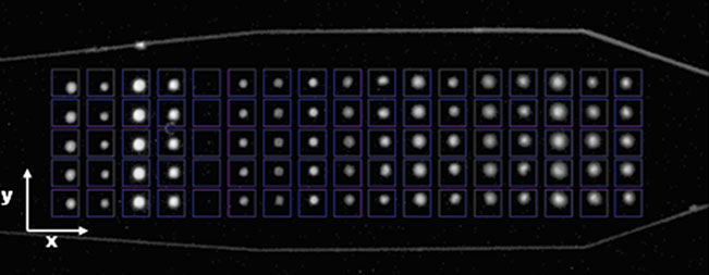
used a grid pattern (see Fig. ), where size and distances of
the other spots of the same analyte or missing of the whole
the quadratic grid cells are adjustable to the spotting array.
spot. This phenomenon can be explained by the existence
For our investigations we created squares with a size of
of small bubbles in the measurement flow channel during
25×25 pixels (equivalent to an area of ca. 1 mm2). In
light exposure. The occurrence of air bubbles in the flow-
relation to the spot diameters, which vary in dependency on
injection system could not be completely excluded, since in
the analyte between 7 and 13 pixels, these cells are large-
this study we did not use any air trap (see Supp. Fig.
scaled. The advantage of the big sized grid cells is the
Thus, an efficient detection of outlier spots before evalua-
entire registration of each spot even if there are drifts in the
tion of the analyte CL intensities is needed to minimize
spotting array, what means that the spots are not immobi-
irregular influences on the dose–response measurements.
lized in perfect horizontal or vertical lines.
For this purpose, the algorithm implemented works on basis
For calculation of the CL signal of one spot the software
of the standard deviations. The first calculation step is the
detects the ten brightest pixels within the corresponding
determination of the overall relative standard deviation (R.
square. The average value represents the CL signal of one
S.D.). Outliers are defined by a certain R.S.D. limit value.
single spot. Defective pixels with grey level intensities in
For our investigations this limit was set to 20%. Exceeding
the saturation region are filtered by an implemented
of the limit value indicates the occurrence of one or more
threshold to exclude artificial influences on the mean value.
outlier spots in the measured analyte column. Because of
Fig. 1 Grid pattern for theevaluation of the chemilumines-cence intensities (differentanalytes in x-direction, repli-cates of the same analyte iny-direction). Size of the gridcells is 25×25 pixels
K. Wutz et al.
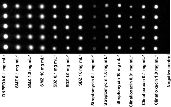
Fig. 2 Characteristic imageof the antibiotic microarray.
Immobilization in three differentspotting concentrations perderivative
this, the second calculation step is the determination of the
With this method outlier spots can be effectively
R.S.D. for the possible combinations x following Eq.
eliminated by choosing the combination of spots without
the outlier. The working principle of the algorithm is
explained in Supp. Table The algorithm developed
allows the elimination of a second outlier per analyte in
In this equation n represents the number of spots per
rare cases of exceeding the limit value though. All
analyte and k is the number of chosen spots for the R.S.D
combinations of three spots (n=5, k=3) are evaluated by
their corresponding R.S.D. and the combination with
Fig. 3 Dose–response curvesfor the four antibiotic analytesenrofloxacin (a), SDZ (b), SMZ(c) and streptomycin (d) inhoney samples. Standard devia-tion is represented by error bars(m=5)
Simultaneous determination of four antibiotic residues in honey
Table 1 Characteristics of the dose–response curves
lization process. For SMZ and SDZ all spotting concen-trations were applicable for the further experiments regarding
IC50 [μg kg−1]
the obtained SNR, but regeneration studies showed strongdetaching effects of excessive molecules on the spot surface
in case of high spotting concentrations (data not shown).
Thus, the spotting concentrations of the antibiotic derivatives
selected for calibration were 0.1 mg mL−1 for SMZ and
clinafloxacin and 1.0 mg mL−1 for SDZ and streptomycin,respectively.
minimal deviation is chosen for calculation of the analyte
Simultaneous dose–response measurements
and determination of recovery
Spotting adjustment for antibiotic microarrays
Dose–response measurements were carried out for thesulfonamides sulfadiazine (SDZ) and sulfamethazine
The establishment of an appropriate microarray immunoas-
(SMZ), the aminoglycoside streptomycin and the fluoro-
say for the detection of antibiotic contaminants requires
quinolone enrofloxacin. The multianalyte ELISA assay on
adjustment of the spotting concentrations. On the one hand,
the microarray chips was performed in an indirect compet-
the amount of analyte molecules immobilized on the spot
itive format, which is most applicable for small analytes.
surface affects the obtained CL signal and has to be
The first two measurements with one chip were used for
optimized to achieve high signal-to-noise ratios (SNR). On
complete loading of reagents to obtain maximal CL signal,
the other hand, the spotting concentration influences the IC50
the third measurement was used for the blank measurement
values of the standard calibration curves and for this reason
with a honey sample without antimicrobial additives.
the working ranges. Furthermore, choosing to high spotting
Afterwards, the standard solutions were measured along
concentrations can lead to an excess of analyte molecules on
increasing analyte concentration. Figure illustrates the
the spot surface detectable by a decrease of the CL signal
resulting dose–response curves. The characteristic standard
during the first measurement cycles due to detaching effects.
calibration data for the four determined analytes are shown
Therefore, different concentrations of the four antibiotics
in Table The working ranges (WR) were defined as
examined were tested for immobilization carrying out blank
10%–80% of the maximum CL signal in case of enroflox-
measurements. The resulting microarray image is depicted in
acin, for the other analytes as 20%–80%.
Fig. the spotting sizes and SNR values are shown in Supp.
For determination of the recovery rates, spiking experi-
ments were carried out. Honey samples were prepared
For all immobilized antibiotic derivatives similar trends
with various contents of the four antibiotics. Spiked
were obtained. With increasing spotting concentrations
samples were directly measured after calibration on the
enhanced CL signal values could be observed. In case of
microarray chip. Due to an observed signal decrease along
streptomycin, the spotting concentration of 0.1 mg mL−1
chip regeneration cycles a blank measurement was done
showed low SNR of 16 : 1 for the blank measurement and
before analysis of each spiked sample. The obtained CL
inhomogeneous surface covering densities, whereas higher
signal (SCL) of the sample measurement was corrected
amounts of streptomycin lead to sufficient CL signals of the
following Eq. To confirm the feasibility of this
spots. The spots measured for the clinafloxacin spotting
correction method the determination of the recovery rates
solution with 0.01 mg mL−1 gave small spot sizes
was performed as duplicates for each sample on the same
compared with the other tested solutions for the immobi-
chip. In addition, a contaminated honey sample that was
Table 2 Recoveries of the spiking experiments
Contaminated sample
1.0 μg kg−1 [μg kg−1]
10 μg kg−1 [μg kg−1]
100 μg kg−1 [μg kg−1]
(25.5 μg kg−1 streptomycin) [μg kg−1]
K. Wutz et al.
determined to 25.5 μg kg−1 streptomycin by LC-MS-MS
milk which need no dilution steps. Due to the regenerability
analysis was examined. The results of the spiking experi-
of the antibiotics microarray each chip could be individu-
ments are presented in Table
ally calibrated before the analysis is performed and more
The dose–response curve for enrofloxacin showed good
than 40 analyses could be done per chip which reduces the
sensitivity with distinct differences of the CL signal in the
costs per analysis and achieve an automated work flow in
range between 1 and 29 μg kg−1, so precise analysis of
routine laboratories. Spiking experiments showed with a
samples within this contamination level is possible. The
recovery rate of ±10% a high accuracy of enrofloxacin
calibrations for SDZ, SMZ and streptomycin dynamic
down to a concentration of 1 μg kg−1. Sulfamethazine,
ranges allow determination of antibiotic contamination
sulfadiazine and streptomycin have a recovery rate between
within a broader span.
75% and 146% at 100 μg kg−1 as this concentration lies in
A high precision of the calibration for enrofloxacin could
the dynamic ranges of their calibration curves in honey.
be confirmed by recovery for the samples spiked with
Finally, the multianalyte immunoassay has successfully
1.0 μg kg−1 and 10 μg kg−1, respectively. The spiked
identified a streptomycin contaminated honey product
enrofloxacin sample of 10 μg kg−1 was in the working
which was approved through LC-MS/MS.
range of the multianalyte immunoassay. A recovery of
Additionally, a new data processing method for CL
92%±6% was achieved. The calculated detection limit was
microarrays images was examined in this study for rapid
4.2 μg kg−1 although a concentration of 1.0 μg kg−1
microarray analysis in routine laboratories. Each spot was
enrofloxacin could be precisely quantified with recovery of
automatically evaluated; outlier could be identified and
95%±7%. This indicates that the precision of this system
were excluded for the analysis. This algorithm is important
depends mainly on the spot quality of the microarray. These
to reduce the variances in the CL-MIA and allows an
differences between the LOD and the analytical sensitivity
automated data processing for analysis.
are also known in other immunoassay assay platforms [
We have shown with this study that a multianalyte
. A concentration of 100 μg kg−1 enrofloxacin was
immunossay based on an automated flow-through chemi-
outside of the working range and therefore, the two
luminescence microarray technique is suitable for the
determined recoveries were 56% and 76%. Thus, samples
quality control of food sample even for such difficult
with high enrofloxacin contamination levels have to be
matrices like honey. Further investigations have to be
diluted before analysis. SDZ and streptomycin could be
focused on the analytical requirements associated with
quantified precisely at 10 μg kg−1 and 100 μg kg−1 with
2002/657/EC for validation as effective screening method
recoveries between 75% and 114%. SMZ shows an over-
for routine residue analysis. In particular, more samples
estimation. The measurement of a honey sample spiked
have to be analyzed to determine the false non-compliant
with 100 μg kg−1 sulfamethazine showed recoveries
rate (α-error) and the false compliant rate (β-error).
of 130% ±21%. Streptomycin had a detection limit of15.9 μg kg−1. Nevertheless, a spiked concentration of
This work was supported by the Bayerische
Forschungsstiftung (BFS AZ-842-08). We would also thank Prof. E.
10 μg kg−1 could be quantified with a recovery of 103%±
Märtlbauer and Dr. R. Dietrich (Lehrstuhl für Hygiene und
9%. The recovery of streptomycin in a contaminated real
Technologie der Milch, LMU München, Oberschleißheim, Germany)
honey sample was 130.4% ± 0.6% referring to the result of
for providing the monoclonal antibodies to enrofloxacin, SDZ, SMZ
the LC-MS/MS analysis which is comparable to other
and streptomycin. Further, we thank Dr. C. Hinkel (Bayerisches Land-esamt für Gesundheit und Lebensmittelsicherheit, Erlangen, Germany)
for the honey samples and Huntsman Corporation (Rotterdam, TheNetherlands) for the free DAPEG samples.
With this study we have shown the first time that the flow-through microarray technique can be used for the rapid
1. Bogdanov S (2006) Contaminants of bee products. Apidologie
analysis of antibiotics in honey. A chemiluminescence
multianalyte chip immunoassay could be performed to
2. Williams DL (2000) A veterinary approach to the european honey
bee (apis mellifera). Vet J 160:61–73
analyse directly antibiotics in honey. No extraction method
3. Hansen H, Brodsgaard CJ (1999) American foulbrood: a review
has to be conducted which simplifies the analysis of honey
of its biology, diagnosis and control. Bee World 80:5–23
samples. Four relevant antibiotics for honey (enrofloxacin,
4. Michaud V (2005) Antibiotic residues in honey - the FEEDM
streptomycin, sulfamethazine and sulfadiazine) could be
view. Apiacta 40:52–54
5. Eastgate JA (2000) Erwinia amylovora: the molecular basis of
quantified simultaneously in 8 min. As result of the high
fireblight disease. Mol Plant Pathol 1:325–329
viscosity of honey, samples were diluted 1:10, leading to
6. Vidaver AK (2002) Uses of antimicrobials in plant agriculture.
reduction of sensitivity compared to other food samples like
Clin Infect Dis 34:S107–S110
Simultaneous determination of four antibiotic residues in honey
7. Wang J (2004) Determination of five macrolide antibiotic residues
simultaneous detection of antibiotics in milk. Anal Chem
in honey by LC-ESI-MS and LC-ESI-MS/MS. J Agr Food Chem
23. Nagl S, Schaeferling M, Wolfbeis OS (2005) Fluorescence
8. Sheridan R, Policastro B, Thomas S, Rice D (2008) Analysis and
analysis in microarray technology. Microchim Acta 151:1–21
occurrence of 14 sulfonamide antibacterials and chloramphenicol
24. Seidel M, Niessner R (2010) Chemiluminescence to immuno-
in honey by solid-phase extraction followed by LC/MS/MS
assays. In: Roda A (ed) Chemiluminescence and bioluminescence.
analysis. J Agr Food Chem 56:3509–3516
R Soc Chem, Cambridge, pp 291–335
9. Pena A, Pelantova N, Lino CM, Silveira MIN, Solich P (2005)
25. Heyries KA, Loughran MG, Hoffmann D, Homsy A, Blum LJ,
Validation of an analytical methodology for determination of
Marquette CA (2008) Microfluidic biochip for chemiluminescent
oxytetracycline and tetracycline residues in honey by hplc with
detection of allergen-specific antibodies. Biosens Bioelectron
fluorescence detection. J Agr Food Chem 53:3784–3788
10. Lopez MI, Pettis JS, Smith IB, Chu PS (2008) Multiclass
26. Schaferling M, Nagl S (2006) Optical technologies for the read
determination and confirmation of antibiotic residues in honey
out and quality control of DNA and protein microarrays. Anal
using LC-MS/MS. J Agr Food Chem 56:1553–1559
Bioanal Chem 385:500–517
11. Hammel YA, Mohamed R, Gremaud E, LeBreton MH, Guy PA
27. Roda A, Guardigli M, Michelini E, Mirasoli M, Pasini P (2003)
(2008) Multi-screening approach to monitor and quantify 42
Analytical bioluminescence and chemiluminescence. Anal Chem
antibiotic residues in honey by liquid chromatography-tandem
mass spectrometry. J Chromatogr A 1177:58–76
28. Tsunoda M (2009) Chemiluminescence detection with separation
12. Debayle D, Dessalces G, Grenier-Loustalot MF (2008) Multi-
techniques for bioanalytical applications. Bioanal Rev 1:25–34
residue analysis of traces of pesticides and antibiotics in honey by
29. Hartwell SK, Grudpan K (2010) Flow based immuno/bioassay
HPLC-MS-MS. Anal Bioanal Chem 391:1011–1020
and trends in micro-immuno/biosensors. Microchim Acta
13. Heering W, Usleber E, Dietrich R, Martlbauer E (1998)
Immunochemical screening for antimicrobial drug residues in
30. Ligler FS (2009) Perspective on optical biosensors and integrated
commercial honey. Analyst 123:2759–2762
sensor systems. Anal Chem 81:519–526
14. Pastor-Navarro N, Gallego-Iglesias E, Maquieira A, Puchades R
31. Langer V, Niessner R, Seidel M (2011) Stopped-flow microarray
(2007) Development of a group-specific immunoassay for
immunoassay for the detection of viable E. coli by use of
sulfonamides - application to bee honey analysis. Talanta
chemiluminescence flow-through microarrays. Anal Bioanal
Chem 399:1041–1050
15. Adrian J, Pinacho DG, Granier B, Diserens JM, Sanchez-Baeza F,
32. Kloth K, Niessner R, Seidel M (2009) Development of an open
Marco MP (2008) A multianalyte ELISA for immunochemical
stand-alone platform for regenerable automated microarrays.
screening of sulfonamide, fluoroquinolone and beta-lactam anti-
Biosens Bioelectron 24:2106–2112
biotics in milk samples using class-selective bioreceptors. Anal
33. Kloth K, Rye-Johnsen M, Didier A, Dietrich R, Martlbauer E,
Bioanal Chem 391:1703–1712
Niessner R, Seidel M (2009) A regenerable immunochip for the
16. Seidel M, Niessner R (2008) Automated analytical microarrays: a
rapid determination of 13 different antibiotics in raw milk.
critical review. Anal Bioanal Chem 391:1521–1544
Analyst 134:1433–1439
17. Valdés M, Valdés González A, García Calzón J, Díaz-García M
34. Dietrich R, Usleber E, Martlbauer E (1998) The potential of
(2009) Analytical nanotechnology for food analysis. Microchim
monoclonal antibodies against ampicillin for the preparation of a
multi-immunoaffinity chromatography for penicillins. Analyst
18. Donhauser SC, Niessner R, Seidel M (2009) Quantification of
E. coli DNA on a flow-through chemiluminescence microarray
35. Wolter A, Niessner R, Seidel M (2007) Preparation and
readout system after pcr amplification. Anal Sci 25:669–674
characterization of functional poly(ethylene glycol) surfaces
19. Magliulo M, Simoni P, Guardigli M, Michelini E, Luciani M, Lelli
for the use of antibody microarrays. Anal Chem 79:4529–
R, Roda A (2007) A rapid multiplexed chemiluminescent immu-
noassay for the detection of Escherichia coli 0157: H7, yersinia
36. Jongberg S, Orlien V, Skibsted LH, Weigel S (2009) Elimination
enterocolitica, salmonella typhimurium, and listeria monocytogenes
of matrix interferences in biosensor analysis of streptomycin in
pathogen bacteria. J Agr Food Chem 55:4933–4939
honey. Eur Food Res Technol 228:659–664
20. Wolter A, Niessner R, Seidel M (2008) Detection of Escherichia
37. Findlay JWA, Dillard RF (2007) Appropriate calibration curve
coli 0157: H7, salmonella typhimurium, and legionella pneumo-
fitting in ligand binding assays. AAPS J 9:E260–E267
phila in water using a flow-through chemiluminescence micro-
38. Albrecht C, Kaeppel N, Gauglitz G (2008) Two immunoassay
array readout system. Anal Chem 80:5854–5863
formats for fully automated CRP detection in human serum. Anal
21. Taitt CR, Shriver-Lake LC, Ngundi MM, Ligler FS (2008) Array
Bioanal Chem 391:1845–1852
biosensor for toxin detection: continued advances. Sensors
39. Petersen AB, Gudmann P, Milvang-Gronager P, Morkeberg R,
Bogestrand S, Linneberg A, Johansen N (2004) Performance
22. Knecht BG, Strasser A, Dietrich R, Martlbauer E, Niessner R,
evaluation of a specific IgE assay developed for the advia centaur
Weller MG (2004) Automated microarray system for the
((r)) immunoassay system. Clin Biochem 37:882–892
Source: http://www.gwk-munich.de/MCR3_14jan11.pdf
Intellectual Property Updates KDN NO.: PP12637/08/2013(032554) Issue #2, August 2012 "Brunei joins the Paris Convention effective CONTENTS: "New Patent Law in Brunei from 17 February 2012" ∙ All Change for Brunei Patents 01 January 2012" ∙ Voluntary Notification
Proposal of a Methodology for Implementing a Service-Oriented Architecture in Distributed I. Medina Buloa,, A. García Domíngueza, F. Aguayob, L. Sevillac and M. Marcosd aDepartment of Computer Languages and Systems. University of Cádiz. School of Engineering. c/ Chile 1, 11002, Cádiz. bDepartment of Design Engineering. University of Seville. Polytechnic University School. c/ Virgen de