Viagra gibt es mittlerweile nicht nur als Original, sondern auch in Form von Generika. Diese enthalten denselben Wirkstoff Sildenafil. Patienten suchen deshalb nach viagra generika schweiz, um ein günstigeres Präparat zu finden. Unterschiede bestehen oft nur in Verpackung und Preis.
Induction of tumour immunity by targeted inhibition of nonsense-mediated mrna decay
Vol 465 13 May 2010
Induction of tumour immunity by targeted inhibitionof nonsense-mediated mRNA decay
Fernando Pastor1, Despina Kolonias1, Paloma H. Giangrande2 & Eli Gilboa1
The main reason why tumours are not controlled by the immune
antigenicity of disseminated tumours leading to their immune
system is that, unlike pathogens, they do not express potent
recognition and rejection. The cell-free chemically synthesized
tumour rejection antigens (TRAs). Tumour vaccination aims at
oligonucleotide backbone of aptamer–siRNAs reduces the risk of
stimulating a systemic immune response targeted to, mostly weak,
immunogenicity and enhances the feasibility of generating
antigens expressed in the disseminated tumour lesions. Main chal-
reagents suitable for clinical use.
lenges in developing effective vaccination protocols are the iden-
Disseminated metastatic disease is the primary cause of death
tification of potent and broadly expressed TRAs1–3 and effective
among cancer patients. Cancer vaccination stimulates a systemic
adjuvants to stimulate a robust and durable immune response4–6.
immune response against judiciously chosen tumour antigens
Here we describe an alternative approach in which the expression
expressed in the tumour cells that seeks out and destroys the disse-
of new, and thereby potent, antigens are induced in tumour
minated tumour lesions. The development of effective cancer vac-
cells by inhibiting nonsense-mediated messenger RNA decay
cines will require the identification of potent and broadly expressed
(NMD)7–10. Small interfering RNA (siRNA)-mediated inhibition
TRAs1–3 as well as effective adjuvants to stimulate a robust and dur-
of NMD in tumour cells led to the expression of new antigenic
able immune response4–6. An alternative approach to vaccination is
determinants and their immune-mediated rejection. In subcutan-
to express new, and hence potent, antigens in tumour cells in situ.
eous and metastatic tumour models, tumour-targeted delivery of
How to express new antigens in the disseminated tumour lesions, but
NMD factor-specific siRNAs conjugated to oligonucleotide apta-
not in normal tissue, have precluded the development of such strat-
mer ligands led to significant inhibition of tumour growth that
egies so far. NMD is an evolutionarily conserved surveillance mech-
was superior to that of vaccination with granulocyte–macrophage
anism in eukaryotic cells that prevents the expression of mRNAs
containing a premature termination codon (PTC)8–10. Inhibition of
tumour cells11, and could be further enhanced by co-stimulation.
NMD in cultured human cell lines using siRNAs targeted to any of its
Tumour-targeted NMD inhibition forms the basis of a simple,
factors, SMG1, UPF1, UPF2 or UPF3, results in the upregulation of
broadly useful, and clinically feasible approach to enhance the
several products encoded by the PTC-containing mRNAs (see, for
Upf2 shRNA
Smg1 shRNA
umour volume (mmT
c )3 1,500
Upf2 shRNA
Smg1 shRNA
centage Pmel-1 per
shRNA
Smg1
shRNA
Smg1
Figure 1 Expression of Upf2 or Smg1 shRNA in CT26 tumour cells leads to
were injected with either OT-I or Pmel-1 transgenic CD81 T cells (three mice
immune-mediated inhibition of tumour growth. a, Intratumoral
per group). Six days later, tumours were excised and analysed for OT-I and
accumulation of OVA-specific OT-I T cells in response to NMD inhibition.
Pmel-1 T-cell content by flow cytometry. Ctrl, control. n 5 2 b, Balb/c mice
B16/F10 tumour cells transduced with shRNA-encoding lentiviral vectors
were implanted subcutaneously with CT26 tumour cells stably transduced
(described in Supplementary Fig. 1a) were stably transfected with an NMD
with the shRNA inducible lentiviral vector encoding Smg1, Upf2 and control
reporter plasmid (described in Supplementary Fig. 1b) containing the class
shRNA (ten mice per group). Each group was divided into two subgroups
I-restricted epitope of chicken ovalbumin (OVA). Mice were implanted
receiving (filled circles) or not receiving (open circles) doxycycline in the
subcutaneously with parental tumour cells (wild-type (WT) B16) or with the
drinking water. n 5 2. c, Same as b except that tumour cells were injected
lentivirus-transduced tumour cells, and either received or did not receive
into immune-deficient nude mice. n 5 1.
doxycycline in their drinking water. When tumours became palpable, mice
1Department of Microbiology & Immunology, Dodson Interdisciplinary Immunotherapy Institute, University of Miami Miller School of Medicine Miami, Florida 33134, USA.
2Department of Internal Medicine and Department of Radiation Oncology, Molecular and Cellular Biology Program, University of Iowa, Iowa City, Iowa 52242, USA.
2010
Macmillan Publishers Limited. All rights reserved
NATURE Vol 465 13 May 2010
example, refs 12–15). Many of these products, resulting from aber-
No T-cell responses were detected against tumour cells that did not
rant splicing or NMD-dependent autoregulated alternative splic-
express Smg1 shRNA or against normal tissues including liver, colon
ing7,8,16, encode new peptides that have not induced tolerance (see
and prostate (Supplementary Fig. 3). This is consistent with the
Supplementary Discussion). We proposed that the upregulation of
hypothesis that tumour rejection was mediated by the induction of
such products when NMD is inhibited in tumour cells will elicit an
immune responses against NMD-controlled products that were
immune response against (some of) the new products, and that the
upregulated when NMD was inhibited in the tumour cells.
immune response will inhibit tumour growth. Moreover, there is
In the experiment shown in Fig. 1b, tumour growth was comple-
evidence that frameshift mutations in cancer cells exhibiting DNA
tely prevented when NMD was inhibited in all tumour cells from the
mismatch repair generate PTC-containing transcripts that are nega-
time of tumour implantation. Simulating a more relevant clinical
tively controlled by NMD17. Inhibiting NMD could, therefore, fur-
model, we tested whether inhibition of NMD in pre-existing tumours
ther augment the production of such tumour-specific antigens (see
can induce therapeutically useful tumour immunity. To preclude
NMD inhibition in normal cells, the NMD factor siRNAs were tar-
To determine whether NMD inhibition in tumour cells can
geted to tumour cells using oligonucleotide aptamer ligands21,22.
stimulate protective anti-tumour immunity, we tested whether
Smg1 and Upf2 siRNA were conjugated to an oligonucleotide apta-
the stable expression of NMD factor short hairpin RNAs
mer that binds to prostate-specific membrane antigen (PSMA)23 as
(shRNAs) in tumour cells inhibits their growth potential in mice.
shown in Supplementary Fig. 4. PSMA-expressing CT26 and B16
CT26 colon carcinoma tumour cells were transduced with a lenti-
tumour cell lines were generated by transduction with a PSMA-
viral vector (PTIG-U6tetOshRNA) encoding Smg1 or Upf2 shRNAs
encoding expression vector, and PSMA expression was confirmed
expressed from a tet-regulated U6 promoter18. shRNA expression
by flow cytometry (not shown). The PSMA-conjugated siRNAs
can be upregulated in vitro by adding doxycycline to the culturemedium, and in vivo by providing doxycycline in the drinkingwater. Doxycycline-induced Smg1 and Upf2 shRNA expression in
cultured CT26 cells results in downregulation of the corresponding
mRNA (Supplementary Fig. 1a) and inhibition of NMD
(Supplementary Fig. 1b). Long-term inhibition of NMD, or other
functions controlled by SMG1 or UPF2, had no measurable effects
Normal lung weight
on the viability or proliferative capacity of the CT26 cells in vitro
(data not shown).
To determine whether siRNA inhibition of NMD in the tumour-
Lung weight (g) 0.5
bearing mice can stimulate immune responses against products that
umour volume (mmT
are normally under NMD control, we measured the intratumoral
accumulation of T cells recognizing a model tumour antigen that is
suppressed as a result of NMD. B16/F10 tumour cells containing thedoxycycline-inducible Smg1, Upf2 or control shRNA were stably
PSMA-ctrl + 4-1BB
transfected with an NMD reporter plasmid encoding the dominantmajor histocompatibility complex (MHC) class I epitope of the
chicken ovalbumin gene (OVA) upstream of a PTC (diagrams in
Fig. 1a and Supplementary Fig. 1a). Tumour-bearing mice wereinfused with OT-I transgenic CD81 T cells that recognize the OVA
MHC class I-restricted epitope20, or with Pmel-1 transgenic CD81 T
cells that recognize an MHC class I-restricted epitope in the endo-genous gp100 tumour antigen expressed in B16 tumour cells19. gp100
expression is not under NMD control. As shown in Fig. 1a, unlikePmel-1 T cells, the OT-I T cells failed to accumulate to significant
PSMA-
Smg1 + mut4-1BB
PSMA-
Smg1 + 4-1BB
levels in the OVA-negative B16/F10 tumours or in tumours trans-
umour volume (mmT
fected with the PTC-containing b-globin-OVA construct encoding
but not expressing Smg1 or Upf2 shRNA. However, upregulation ofSmg1 or Upf2 shRNA, but not control shRNA (doxycycline in the
drinking water) resulted in a significant accumulation of OT-I T cells
in the tumours. This experiment shows that siRNA inhibition of
NMD in tumour cells can induce an immune response in vivo againstan antigen that is under NMD control.
To determine whether siRNA-mediated inhibition of NMD affects
tumour growth, the lentiviral-transduced CT26 cells expressing a
Figure 2 Inhibition of tumour growth in mice treated with PSMA aptamer
control, Smg1 or Upf2 shRNA were implanted subcutaneously into
targeted Upf2 and Smg1 siRNAs. a, Balb/c mice were implanted
mice and tumour growth was monitored in the presence or absence
subcutaneously with PSMA-CT26 tumour cells and 3 days later injected via
of doxycycline administered in the drinking water. Figure 1b shows
the tail vein with PBS (filled circles) or with PSMA aptamer–siRNA
that tumour cells expressing Smg1 or Upf2 shRNA, but not control
conjugates (open circles, control siRNA; open squares, Upf2 siRNA; filled
shRNA, grew initially but failed to progress. Tumour inhibition was
squares, Smg1 siRNA) (5 mice per group). n 5 2. b, C57BL/6 mice were
immune-mediated because the tumours grew in nude mice (Fig. 1c),
implanted with PSMA-B16/F10 tumour cells by tail vein injection, and
and mice that rejected the tumours shown in Fig. 1b, but not age-
5 days later were injected with PSMA aptamer–siRNA conjugates (ten miceper group). Metastatic load was determined by measuring lung weight at the
matched control mice, resisted a second challenge with parental
time of euthanization. n 5 2. c, Combination immunotherapy using NMD
tumour cells (not shown). Delaying doxycycline treatment of mice
inhibition and 4-1BB co-stimulation. PSMA-CT26 tumour-bearing mice
expressing Smg1 shRNA diminished the tumour inhibitory effect that
(five mice per group) were treated with various combinations of PSMA
was completely lost when drug treatment was delayed for 6 days
aptamer conjugated to Smg1 or control siRNA and an agonistic or co-
(Supplementary Fig. 2). Tumour rejection correlated with the induc-
stimulation-deficient 4-1BB aptamer dimer26 (mut4-1BB) and monitored
tion of T-cell responses against tumour cells expressing Smg1 shRNA.
for tumour growth. n 5 1.
2010
Macmillan Publishers Limited. All rights reserved
NATURE Vol 465 13 May 2010
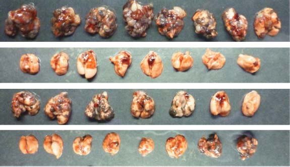
Figure 3 PSMA aptamer–Smg1 siRNA rejectionof PSMA-expressing, but not parental, CT26tumour cells. a
, Mice were co-implanted
subcutaneously with PSMA-expressing (leftflank) and parental (right flank) CT26 tumour
cells and injected with PSMA aptamer–Smg1
siRNA via the tail vein. b, Fifteen days after
tumour inoculation, 32P-labelledaptamer–siRNA was injected, and 3 or 24 h later
tumours were excised and the 32P content
determined. n 5 3. c, Three days after tumour
PSMA-Smg1 siRNA
inoculation, mice were injected with
aptamer–siRNA conjugate (eight mice per group)
as described in Fig. 2a and tumour growth was
Smg1 siRNA
monitored. Open circles, parental CT26; filled
circles, PSMA-CT26. n 5 2.
umour volume (mmT
bound to and were taken up by PSMA-expressing, but not parental,
6. As shown in Fig. 2c, combination therapy with PSMA aptamer–
tumour cells (Supplementary Fig. 5), leading to the downregulation
Smg1 siRNA and 4-1BB aptamer was more than additive.
of their target RNAs (Supplementary Fig. 6).
To determine whether tumour inhibition shown in Fig. 2 is a result
We next tested whether systemic administration of PSMA apta-
of aptamer targeting of siRNA to PSMA-expressing tumour cells, mice
mer–siRNA conjugates by tail vein injection can inhibit tumour
were implanted in opposite flanks with PSMA-expressing and parental
growth. As shown in Fig. 2a, treatment of day 3 subcutaneously
CT26 tumour cells and PSMA aptamer conjugated to control or Smg1
implanted PSMA-CT26 tumour cells with PSMA-conjugated Smg1
siRNA was administered systemically by tail vein injection (Fig. 3a).
siRNA, and to a lesser extent Upf2 siRNA, significantly inhibited
Figure 3b shows that 32P-labelled PSMA aptamer–Smg1 siRNA con-
tumour growth. Two out of seven mice treated with the PSMA apta-
jugate accumulated preferentially in PSMA-expressing tumour cells.
mer–Smg1 siRNA conjugate rejected the implanted tumours and
Figure 3c shows that systemic administration of PSMA aptamer-con-
remained tumour-free (Supplementary Fig. 7). When treatment
jugated Smg1, but not control, siRNA inhibited the growth of PSMA-
intensity was increased by doubling the dose of the aptamer–
expressing CT26 tumour cells but not the contralaterally implanted
siRNA conjugate and extending treatment to seven injections, six
parental CT26 tumour cells. Supplementary Fig. 8 shows a snapshot of
out of seven of mice rejected the tumour long term. Treatment with
the tumour-bearing mice at the day of euthanization.
PSMA aptamer conjugated to control siRNA had a small inhibitory
To assess the potency of tumour-targeted NMD inhibition, we
effect that could have resulted from the binding of the PSMA apta-
compared the anti-tumour effects of treating tumour-bearing mice
mer–siRNA to the tumour cells, or be due to non-specific immune
with PSMA aptamer–Smg1 siRNA conjugate and vaccination with
stimulatory effects of the oligonucleotide24,25. We found no increase
GM-CSF-expressing irradiated syngeneic tumour cells (GVAX)11,27.
in IFNa levels in the serum of mice treated with PSMA aptamer–
In therapeutic protocols when vaccination is initiated 2–4 days
control or Smg1 siRNA conjugates (data not shown). As shown in
after tumour inoculation, the anti-tumour impact of GVAX is
Fig. 2b, the treatment of day 5 PSMA-B16/F10 tumour-implanted
limited, unless combined with other treatments such as CTLA-4
mice with PSMA aptamer-conjugated Upf2 or Smg1 siRNA inhibited
blockade28 or T-regulatory cell depletion29. As shown in Fig. 4, in
the development of lung metastasis that was more profound in the
the B16 lung metastasis model described in Fig. 2b, GVAX treat-
SMG1 group. To determine whether the anti-tumour response eli-
ment of day 1 tumour bearing mice significantly inhibited meta-
cited by NMD inhibition can be further enhanced by co-stimulation,
stasis, whereas treatment of day 5 tumour bearing mice had a
PSMA-CT26 tumour-bearing mice were treated with PSMA apta-
limited anti-metastatic effect that barely reached statistical signifi-
mer–Smg1 siRNA and an agonistic 4-1BB aptamer dimer26. The
cance. By comparison, treatment of day-5 tumour-bearing mice
stringency of NMD inhibition and 4-1BB co-stimulation was
with PSMA aptamer–Smg1 siRNAs inhibited metastasis to an
adjusted to elicit a limited anti-tumour effect when applied separately
extent comparable to that of administering GVAX at day 1.
by delaying treatment with PSMA aptamer–siRNA conjugates from
Given that these are first generation aptamer–siRNA conjugates
days 3 to 5 and administering a single dose of 4-1BB aptamer on day
and the dose and schedule of aptamer–siRNA treatment have not
P < 0.0001
Figure 4 Comparison of PSMA aptamer–Smg1siRNA treatment to vaccination with GM-CSF
P = 0.0442
expressing irradiated tumour cells. C57BL/6mice were injected intravenously with B16/F10
P = 0.0012
tumour cells and treated with PSMAaptamer–siRNA conjugates starting at day 5 as
described in Fig. 2b, or vaccinated with GM-CSF-
expressing irradiated B16/F10 tumour cells
PSMA-Smg1 (D5)
(GVAX) starting at days (D) 1 or 5 using the
protocol described previously29. n 5 1.
2010 Macmillan Publishers Limited. All rights reserved
NATURE Vol 465 13 May 2010
been optimized, these results indicate that tumour-targeted siRNA-
¨hlemann, O., Eberle, A. B., Stalder, L. & Zamudio Orozco, R. Recognition and
mediated NMD inhibition is more effective than a commonly used
elimination of nonsense mRNA. Biochim. Biophys. Acta 1779, 538–549 (2008).
Jinushi, M., Hodi, F. S. & Dranoff, G. Enhancing the clinical activity of granulocyte-
macrophage colony-stimulating factor-secreting tumor cell vaccines. Immunol.
Tumour-targeted NMD inhibition is a new approach to stimulate
Rev. 222, 287–298 (2008).
protective anti-tumour immunity. Instead of stimulating or poten-
12. El-Bchiri, J. et al. Nonsense-mediated mRNA decay impacts MSI-driven
tiating immune responses against existing, often weak, antigens
carcinogenesis and anti-tumor immunity in colorectal cancers. PLoS One 3, e2583
expressed in the tumour cells—the goal of current tumour vaccina-
13. Mendell, J. T., Sharifi, N. A., Meyers, J. L., Martinez-Murillo, F. & Dietz, H. C.
tion protocols—NMD inhibition generates new antigenic determi-
Nonsense surveillance regulates expression of diverse classes of mammalian
nants in situ in the disseminated tumour lesions. It should be noted
transcripts and mutes genomic noise. Nature Genet. 36, 1073–1078 (2004).
that NMD control of gene expression is ‘leaky'. In addition to the first
14. Usuki, F. et al. Specific inhibition of nonsense-mediated mRNA decay
round of translation, known as pioneer translation, the efficiency of
components, SMG-1 or Upf1, rescues the phenotype of Ullrich disease fibroblasts.
nonsense-mediated degradation varies among individual mRNA
Mol. Ther. 14, 351–360 (2006).
15. Wittmann, J., Hol, E. M. & Jack, H. M. hUPF2 silencing identifies physiologic
targets8–10. Immune recognition is, therefore, a consequence of upre-
substrates of mammalian nonsense-mediated mRNA decay. Mol. Cell. Biol. 26,
gulation of NMD-controlled products above a certain threshold that
1272–1287 (2006).
was set by the natural immune tolerance mechanisms. The NMD
16. Isken, O. & Maquat, L. E. The multiple lives of NMD factors: balancing roles in
inhibition strategy described in this study is simple, consisting of a
gene and genome regulation. Nature Rev. Genet. 9, 699–712 (2008).
single reagent that can be synthesized by a cell-free chemical process,
17. Duval, A. & Hamelin, R. Mutations at coding repeat sequences in mismatch
repair-deficient human cancers: toward a new concept of target genes for
it obviates the need to identify TRAs or adjuvants, and is broadly
instability. Cancer Res. 62, 2447–2454 (2002).
applicable as it targets a common pathway in all tumours. The
18. Aagaard, L. et al. A facile lentiviral vector system for expression of doxycycline-
potency of the NMD inhibition approach was suggested when com-
inducible shRNAs: knockdown of the pre-miRNA processing enzyme Drosha.
pared to GVAX vaccination. Arguably, these first generation apta-
Mol. Ther. 15, 938–945 (2007).
mer–siRNA conjugates and the dose and treatment schedule can be
19. Overwijk, W. W. et al. Tumor regression and autoimmunity after reversal of a
functionally tolerant state of self-reactive CD81 T cells. J. Exp. Med. 198, 569–580
further optimized. It would be of interest to determine in future
studies whether the NMD-induced antigens are cross-reactive among
20. Hogquist, K. A. et al. T cell receptor antagonist peptides induce positive selection.
different tumours, and if so to identify the dominant antigens
Cell 76, 17–27 (1994).
induced by NMD inhibition.
21. Gold, L. Oligonucleotides as research, diagnostic, and therapeutic agents. J. Biol.
Chem. 270, 13581–13584 (1995).
22. Nimjee, S. M., Rusconi, C. P. & Sullenger, B. A. Aptamers: an emerging class of
therapeutics. Annu. Rev. Med. 56, 555–583 (2005).
Tumour immunotherapy studies. Three-hundred-thousand parental or pTIG-
23. Lupold, S. E., Hicke, B. J., Lin, Y. & Coffey, D. S. Identification and
U6tetOshRNA transduced CT26 tumour cells were implanted subcutaneously in
characterization of nuclease-stabilized RNA molecules that bind human
Balb/c or Nude mice. At the day of tumour implantation, mice started receiving
prostate cancer cells via the prostate-specific membrane antigen. Cancer Res.
water supplemented with 10% sucrose with or without 2 mg ml21 doxycycline
62, 4029–4033 (2002).
24. Akira, S., Uematsu, S. & Takeuchi, O. Pathogen recognition and innate immunity.
To evaluate the anti-tumour effects of PSMA aptamer–siRNAs, mice were
Cell 124, 783–801 (2006).
implanted with 1 3 106 PSMA-CT26 tumour cells and injected with 400 pmoles
25. Judge, A. D. et al. Sequence-dependent stimulation of the mammalian innate
of aptamer–siRNA in 100 ml PBS via the tail vein at days 3, 5, 7, 9, 11 and 13. In
immune response by synthetic siRNA. Nature Biotechnol. 23, 457–462
combination therapy, treatment with PSMA aptamer–siRNA was administered
26. McNamara, J. O. et al. Multivalent 4-1BB binding aptamers costimulate CD8 T
at days 5, 7, 9, 11 and 13, and a single dose of 500 pmoles of 4-1BB aptamer dimer
cells and inhibit tumor growth in mice. J. Clin. Invest. 118, 376–386 (2008).
was administered on day 6.
27. Dranoff, G. et al. Vaccination with irradiated tumor cells engineered to secrete
To monitor metastasis, C57BL/6 mice were implanted with 105 B16-PSMA
murine granulocyte-macrophage colony-stimulating factor stimulates potent,
transduced cells by the tail vein and injected with 400 pmoles of aptamer-
specific, and long-lasting anti-tumor immunity. Proc. Natl Acad. Sci. USA 90,
siRNA conjugates at days 5, 8, 11, 14 and 17. When about half of the mice in
3539–3543 (1993).
the control groups had shown signs of morbidity (approximately days 25–28),
28. van Elsas, A., Hurwitz, A. A. & Allison, J. P. Combination immunotherapy of B16
the mice were euthanized and their lungs were weighed. GM-CSF-expressing
melanoma using anti-cytotoxic T lymphocyte-associated antigen 4 (CTLA-4)
B16/F10 tumour cells, provided by G. Dranoff, were irradiated (50 Gy) and
and granulocyte/macrophage colony-stimulating factor (GM-CSF)-producing
5 3 105 cells were injected subcutaneously at days 1, 4 and 7, or days 5, 8 and
vaccines induces rejection of subcutaneous and metastatic tumors accompanied
11, as described previously29.
by autoimmune depigmentation. J. Exp. Med. 190, 355–366 (1999).
For statistical analysis, P values were calculated using a Student's t-test.
29. Quezada, S. A., Peggs, K. S., Curran, M. A. & Allison, J. P. CTLA4 blockade and
GM-CSF combination immunotherapy alters the intratumor balance of effector
Full Methods and any associated references are available in the online version of
and regulatory T cells. J. Clin. Invest. 116, 1935–1945 (2006).
Supplementary Information is linked to the online version of the paper at
Received 21 October 2009; accepted 2 March 2010.
Acknowledgements We thank J. Zhang for assistance in the mouse studies, A.-M.
Gilboa, E. The makings of a tumor rejection antigen. Immunity 11, 263–270 (1999).
Jegg for technical assistance in characterizing Smg1 siRNAs, J. Rossi for advising in
Novellino, L., Castelli, C. & Parmiani, G. A listing of human tumor antigens
the design of aptamer–siRNA conjugates, and S. Nair and D. Boczkowski for advice in
recognized by T cells. Cancer Immunol. Immunother. 54, 187–207 (2005).
performing T-cell assays. This work was supported by the Dodson foundation and
Schietinger, A., Philip, M. & Schreiber, H. Specificity in cancer immunotherapy.
the Sylvester Comprehensive Cancer Center (Medical School, University of Miami).
Semin. Immunol. 20, 276–285 (2008).
Gilboa, E. The promise of cancer vaccines. Nature Rev. Cancer 4, 401–411 (2004).
Author Contributions F.P. suggested the approach and was responsible for
Melief, C. J. Cancer immunotherapy by dendritic cells. Immunity 29, 372–383 (2008).
designing the aptamer–siRNA conjugates and interpreting the results, D.K. was
Pardoll, D. M. Spinning molecular immunology into successful immunotherapy.
responsible for the mouse studies, P.H.G. helped design the aptamer–siRNA
Nature Rev. Immunol. 2, 227–238 (2002).
conjugates, and E.G. oversaw experimental design, data analysis, and wrote the
Frischmeyer, P. A. & Dietz, H. C. Nonsense-mediated mRNA decay in health and
disease. Hum. Mol. Genet. 8, 1893–1900 (1999).
Behm-Ansmant, I. et al. mRNA quality control: an ancient machinery recognizes
Author Information Reprints and permissions information is available at
and degrades mRNAs with nonsense codons. FEBS Lett. 581, 2845–2853 (2007).
The authors declare no competing financial interests.
Maquat, L. E. Nonsense-mediated mRNA decay: splicing, translation and mRNP
Correspondence and requests for materials should be addressed to E.G.
dynamics. Nature Rev. Mol. Cell Biol. 5, 89–99 (2004).
2010 Macmillan Publishers Limited. All rights reserved
PSMA aptamer–siRNA conjugates. The PSMA aptamer, 59-GGGAGG
CTGAGGAGAAG-39 and reverse primer 59-GGGTGTTGGCGGGTGTC-39,
cloned in the pcDNA3.1 plasmid (Invitrogen) and used to transfect parental
CGGCAGACGACUCGCCCGA-39 was cloned into pUC57 between KpnI
and pTIG-U6tetOshRNA transduced B16/F10 tumour cells.
and BamHI restriction sites. siRNAs were screened using the psiCHECK
RT–PCR. RNA was isolated using RNAsy columns (Qiagen) from cells grown in
system (Promega) from candidates generated by the HPCdispatcher and
the presence or absence of 1 mg ml21 doxycycline (Sigma) for 5 days were
OpenBiosystem algorithms. The DNA template for the aptamer–siRNA guide
reverse-transcribed and PCR-amplified using the following primers. CT26 and
strand was generated by PCR amplification using forward primer 59-
pTIG-U6tetOshRNA transduced CT26 tumour cells: actin: forward, 59-
TAATACGACTCACTATAGGGAGGACGATGCGG-39 and reverse primers
GC-39. Smg1: forward, 59-GCCCATCGTGTTTGCTTTGG-39; reverse, 59-
TCTCGTTCCCAGTGGTGTTACAG-39. Upf2: forward, 59-ACCCGGGGCUA
AUGUUGAC-39; reverse, 59-CUUGGUAAUGUUAGGCGUUUUCUC-39. BG,
for Smg1 siRNA. The PCR products were purified using the QIAprep Spin col-
BGPTC and OVA-BGPTC transduced cells: b-globin: forward, 59-ACCACC
umns (Qiagen) RNA was transcribed using the T7(Y639F) polymerase and hybri-
dized to the corresponding passenger strands (control siRNA sequence: 59-
Transfection of cells with aptamer–siRNA conjugates. CT26 and PSMA-CT26
AAUUCUCCGAACGUGUCACdTdT-39; Upf2 siRNA sequence: 59-GCGUUA
tumour cells were incubated with 400 nM siRNA or PSMA aptamer–siRNA
UGUUUGGUGGAAGdTdT-39; Smg1 siRNA sequence: 59-GCCAUGACUAA
conjugate in the presence of absence of Lipofectamine 2000 (Invitrogen) for
2 days and analysed for RNA expression or NMD inhibition.
Derivation of PSMA-expressing CT26 tumour cell lines. The PSMA comple-
Tumour infiltration of OT-1 and Pmel-1 T cells. C57BL/6 mice (CD45.2;
mentary DNA, provided by V. Ponomarev, was PCR-amplified using forward
Thy1.2) were implanted subcutaneously with 5 3 104 B16 tumour cells and 8
days after tumour inoculation 5 3 106 peptide-activated OT-I (CD45.1) or
Pmel-1 CD81 T cells were injected intravenously via the tail vein. At the same
ACTTCACTC-39, and cloned into the SalI and Not1 restriction sites of the retro-
day the drinking water was supplemented with 10% sucrose (Sigma) and with or
viral vector pBMN (Addgene). Plasmid was transiently transfected into the
without 2 mg ml21 doxycycline (Sigma). At day 14 after tumour implantation
Phoenix-AMPHO 293 packaging cell lines and viral supernatant was used to
mice were euthanized, tumours removed and mechanically disaggregated by
transduce CT26 colon carcinoma (H-2d) and B16/F10 melanoma (H-2b) tumour
collagenase treatment (400 U ml21). Cells were ficolled and stained with
cell lines. PSMA-expressing cells were isolated by cell sorting using PSMA-PE-
FITC-labelled anti-CD45.1 antibody and allophycocyanin (APC)-labelled anti-
labelled anti-PSMA antibody from MBL.
CD8 antibody for OT-1 T cells or with phycoerythrin (PE)-labelled anti-Thy1.1
Confocal microscopy. The passenger strand of the siRNAs was labelled with Cy3
antibody and APC-labelled anti-CD8 antibody for Pmel-1 T cells and analysed
before hybridization to the PSMA-aptamer guide strand using the Silencer RNA
by flow cytometry. All antibodies used were from BD Bioscience.
labelling kit (Ambion). Tumour cells were plated on glass plates, washed with
Tumour homing or 32P-labelled atpamer–siRNA conjugates. The PSMA apta-
PBS and incubated with 40 nM of Cy3-labelled aptamer–siRNA or with
mer was transcribed in vitro in the presence of 1/1,000 parts of a32P-ATP
10 mg ml21 anti-PSMA antibody (MBL) and Alexa Fluor 488 goat anti-mouse
(3000 Ci mmol21) (PerkinElmer) and annealed to Smg1 siRNA as described
IgG (Molecular Probes). Coverslips were mounted with Prolong Gold-DAPI
earlier. Balb/c mice were co-implanted with CT26 and PSMA-CT26 tumour cells
(Molecular Probes).
in the opposite flanks, and 15 days later injected via the tail vein with
Generation of stably transduced shRNA-expressing CT26 and B16/F10
5 3 105 c.p.m. 32P-labelled aptamer–siRNA. After aptamer–siRNA injection,
tumour cell lines. Double-stranded oligonucleotides corresponding to the guide
tumours were surgically removed, cells dispersed by incubation with
and passenger strands of Smg1, Upf2 or control siRNA modified to contain
400 U ml21 of collagenase, washed three times with PBS, and cell-associated32
overhangs compatible with BglII and KpnI restriction sites were cloned into
P was measured in a scintillation counter.
the BglII and KpnI sites of pFRT-U6tetO plasmid30. The U6tetO-shRNA cas-
Tumour immunotherapy studies. Three-hundred-thousand parental or pTIG-
settes from the pFRT plasmids were isolated by PCR (forward primer: 59-
U6tetOshRNA transduced CT26 tumour cells were implanted subcutaneously in
Balb/c or Nude mice. At the day of tumour implantation mice started receiving
59-GTTAAGCATGCCCACACTGGACTAGTGGATC-39) and cloned into the
water supplemented with 10% sucrose with or without 2 mg ml21 doxycycline
NotI/SphI restriction sites of PTIG lentiviral vector to generate pTIG-
U6tetOshRNA plasmids30. pTIG-U6tetOshRNA DNA was cotransfected into
To evaluate the anti-tumour effects of PSMA aptamer–siRNAs, mice were
293T cells with lentiviral packaging plasmids pCHPG-2, pCMV-rev and
implanted with 1 3 106 PSMA-CT26 tumour cells and injected with 400 pmoles
PCMV-gag and lentivirus-containing supernatant was collected and concen-
of aptamer–siRNA in 100 ml PBS via the tail vein at days 3, 5, 7, 9, 11 and 13. In
trated by centrifugation31. CT26 colon carcinoma (H-2d) and B16/F10 mela-
combination therapy, treatment with PSMA aptamer–siRNA was administered
noma (H-2b) tumour cell lines were infected with lentiviral vectors and stably
at days 5, 7, 9, 11 and 13, and a single dose of 500 pmoles of 4-1BB aptamer dimer
transduced GFP-expressing cells were isolated by sorting.
was administered on day 6.
shRNA oliognuclotides used were as follows. Control shRNAs: 59-
To monitor metastasis, C57BL/6 mice were implanted with 105 B16-PSMA
transduced cells via the tail vein and injected with 400 pmoles of aptamer–siRNA
conjugates at days 5, 8, 11, 14 and 17. When about half of the mice in the control
AGGAAGTGACACGTTCGGAGAATT-39. Upf2 shRNA: 59-GATCGCGTTATG
groups had shown signs of morbidity (approximately days 25–28), the mice were
euthanized and their lungs were weighed. GM-CSF-expressing B16/F10 tumour
cells, provided by G. Dranoff, were irradiated (50 Gy) and 5 3 105 cells were
TCCACCAAACATAACGC-39. Smg1 shRNA: 59-GATCGCCACCAAAGACA
injected subcutaneously at days 1, 4 and 7, or days 5, 8 and 11, as described
For statistical analysis P values were calculated using a Student's t-test.
30. Aagaard, L. et al. A facile lentiviral vector system for expression of doxycycline-
CT26 and B16/F10 tumour cell lines containing BG, BGPTC and OVA-BGPTC.
inducible shRNAs: knockdown of the pre-miRNA processing enzyme Drosha.
The SIINFEKL peptide was cloned into the first exon of the b-globin gene
Mol. Ther. 15, 938–945 (2007).
between second (valine) and third (histidine) amino-terminal amino acids of
31. Li, M. J. & Rossi, J. J. Lentiviral vector delivery of recombinant small interfering
the BG and BGPTC plasmids3, provided by L. Maquat, by PCR using the forward
RNA expression cassettes. Methods Enzymol. 392, 218–226 (2005).
2010 Macmillan Publishers Limited. All rights reserved
Source: http://cmb1.auckland.ac.nz/medsci708/Sun/Pastor%20-discussion%20-Induction%20of%20tumour%20immunity%20by%20targeted%20%20%20inhibition%20of%20nonsense-mediated%20mRNA%20decay.pdf
Published Ahead of Print on March 8, 2010 as 10.1200/JCO.2009.24.4798 JOURNAL OF CLINICAL ONCOLOGY Prediction of Risk of Distant Recurrence Using the 21-GeneRecurrence Score in Node-Negative and Node-PositivePostmenopausal Patients With Breast Cancer Treated WithAnastrozole or Tamoxifen: A TransATAC StudyMitch Dowsett, Jack Cuzick, Christopher Wale, John Forbes, Elizabeth A. Mallon, Janine Salter, Emma Quinn,Anita Dunbier, Michael Baum, Aman Buzdar, Anthony Howell, Roberto Bugarini, Frederick L. Baehner,and Steven Shak
Journal of African Studies in Educational Management and Leadership Vol: 7 No:1, August 2016, 61-81 Scholarly, Peer Reviewed Interrogating Social Media Netiquette and Online Safety among University Students from Assorted Disciplines Simon Macharia Kamau, Khadiala Khamasi & Margaret Kamara Kosgey Abstract


