Viagra gibt es mittlerweile nicht nur als Original, sondern auch in Form von Generika. Diese enthalten denselben Wirkstoff Sildenafil. Patienten suchen deshalb nach viagra generika schweiz, um ein günstigeres Präparat zu finden. Unterschiede bestehen oft nur in Verpackung und Preis.
Pii: s0304-3940(02)01465-9
Neuroscience Letters 339 (2003) 49–52
Functional MRI of amblyopia before and after levodopa
Chao-I Yanga, Meng-Ling Yanga, Ju-Chuan Huangb,c, Yung-Liang Wanb,c, Ray Jui-Fang Tsaia,
Yau-Yau Waib,c, Ho-Ling Liub,c,*
aDepartment of Ophthalmology, Chang-Gung Memorial Hospital, Taoyuan, Taiwan
bDepartment of Diagnostic Radiology, Chang-Gung Memorial Hospital, Taoyuan, Taiwan
cSchool of Medical Technology, Chang-Gung University, Taoyuan, Taiwan
Received 23 October 2002; received in revised form 14 December 2002; accepted 16 December 2002
Functional magnetic resonance imaging (fMRI) was applied to five older amblyopes with monocular amblyopia before and after levodopa
treatment. During the experiment, images were acquired in two runs with visual stimulation delivered through the sound and the amblyopiceyes, respectively. The experiment was performed on each of the subjects, before and after their oral administration of levodopa/carbidopa(0.5/0.12 mg/kg) three times per day for 7 weeks. Our study demonstrated that there was no effect on the spatial extent of the visual corticalactivation during the sound eye stimulation (P ¼ 0:17), but some improvement during the amblyopic eye stimulation (P ¼ 0:06). Thevolume ratio between the amblyopic and sound eye stimulation significantly increased after the treatment (P , 0:05). This finding supportsthe previous studies of levodopa effect on amblyopia at the visual cortical level, and suggests that fMRI can be a useful tool in assessingchanges of visual cortical activity after the treatment
q 2003 Elsevier Science Ireland Ltd. All rights reserved.
Keywords: MRI; fMRI; Vision; Amblyopia; Levodopa
Amblyopia is a unilateral or bilateral reduction of best-
amblyopia) and neurotransmitter activity. Leguire et al.
corrected visual acuity that cannot be attributed directly to
reported that contrast sensitivity function was improved in
the effect of any structural abnormality of the eye or the
patients after a single dose and a 7-week regimen of
posterior visual pathway. It is caused by abnormal visual
levodopa, combined with part-time occlusion
experience early in life, usually resulting from ocular
Recently, functional magnetic resonance imaging
misalignment or uncorrected refractive error. Recently,
(fMRI) based on blood oxygenation level-dependent
Gottlob and Stangler-Zuschrott found that a single dose of
(BOLD) contrast has been utilized to measure the neuronal
levodopa, a precursor of the dopamine, with a combination
activity within visual cortical areas in patients with
of a peripheral decarboxylase inhibitor (e.g. benzerazide or
amblyopia BOLD contrast-based imaging utilizes
carbidopa), can temporarily improve contrast sensitivity in
paramagnetic deoxyhemoglobin as an endogenous contrast
the amblyopic eyes in adults The dopamine is presented
agent During functional brain activity, the
in retinal amacrine and interplexiform cells in humans as a
relative change of cerebral metabolic rate of oxygen was
neurotransmitter and is likely involved in the visual
found to be much less than that of cerebral blood flow,
information transmission to the visual cortex . Occlusion
leading to an increase of the BOLD signal. Using fMRI,
of newborn infant monkey's eye decreased retinal
Goodyear et al. reported that there were fewer activated
dopamine concentration and demonstrated an association
voxels, in the visual cortex, during visual stimulation
between functional changes in the visual pathway (e.g.
through the amblyopic eye than that through the normal eyeChoi et al. demonstrated that the fMRI signal changes in
* Corresponding author. MRI Center, Department of Diagnostic
the calcarine activation were smaller when viewed with
Radiology, Chang Gung Memorial Hospital, 5 Fuhsing Street, Kweishan,
amblyopic eyes than with sound eyes . The purpose of the
Taoyuan 333, Taiwan. Tel.: þ 886-3-3281200x8407; fax: þ 886-3-
present study was to apply fMRI techniques in the
E-mail address:
[email protected] (H.-L. Liu).
0304-3940/03/$ - see front matter q 2003 Elsevier Science Ireland Ltd. All rights reserved.
doi:10.1016/S0304-3940(02)01465-9
C.-I Yang et al. / Neuroscience Letters 339 (2003) 49–52
investigation of the changes of visual cortical activity in
acquired during the stimulation states and those during the
patients with amblyopia after the levodopa treatment.
resting states, using an unpaired Student's t-test with a
Five older amblyopes (aged 13 – 17 years, cases 1, 2, and
threshold of 2.4 (P , 0:01). Regions of interest (ROIs) were
5 were anisometropic, and cases 3 and 4 were anisometropic
chosen in the gray matter area along the calcarine fissure
and strabismic) were enrolled in this study. The indications,
and enclosed the primary visual cortex, excluding the
risks and benefits of the procedures were well explained to
sagittal sinus, based on the anatomical images. For each
the patients or their family, and signed consent was obtained
subject, the volume of significantly activated voxels within
from all the parents before the procedure. All patients
the ROIs, were used to compare the neuronal activity
underwent a detailed ophthalmologic examination and were
induced by visual stimulation through the sound eye and the
considered stable amblyopes who were unresponsive to
amblyopic eye, before and after the treatment. To minimize
traditional occlusion treatment. The best-corrected visual
the variation of experimental conditions before and after
acuities (BCVAs) of their sound eye were all 1.0, while that
taking levodopa, the ratio of the activation volumes from the
of their amblyopic eye were 0.3, 0.3, 0.04, 0.3 and 0.3,
amblyopic and sound eye stimulation was calculated to
respectively. All patients received levodopa and part-time
further investigate the treatment effect.
(3 h/day) occlusion of the sound eye. Levodopa, in the
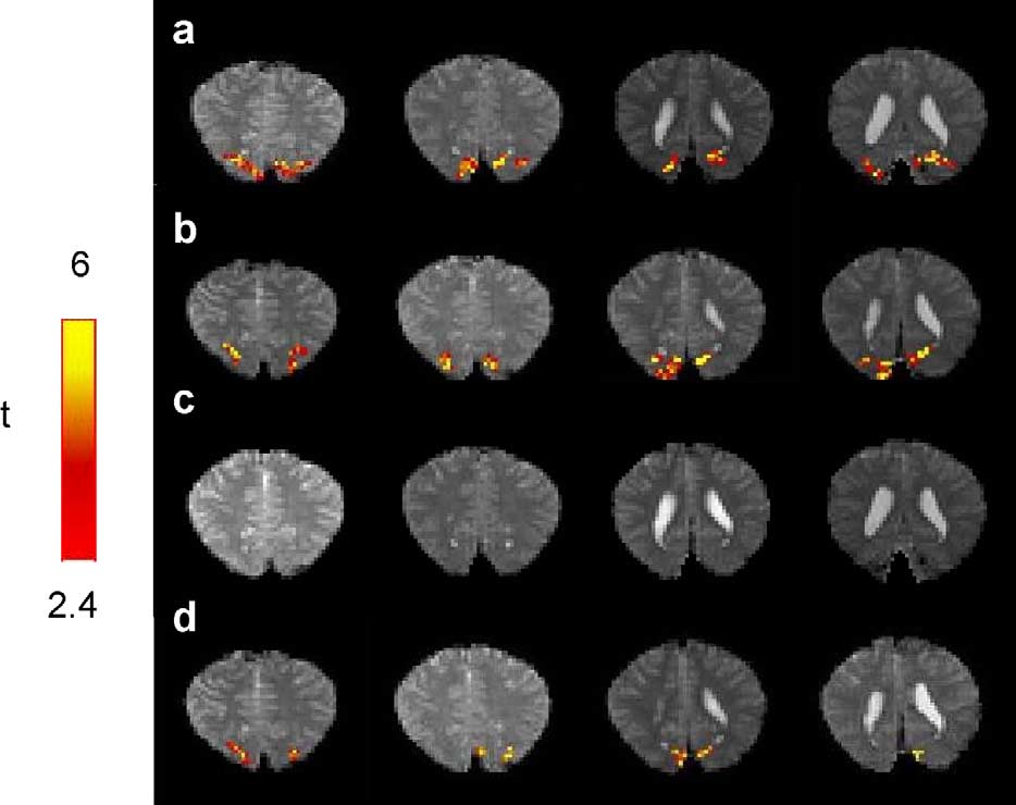
shows the functional maps of visual cortical areas
dosage of 0.5 mg/kg body weight, with a 25% fixed dose
from one of the patients, before and after levodopa
combination of carbidopa was administered orally three
medication. With the stimulation delivered through the
times a day for 7 weeks. The regimen was set according to
sound eye (a,b), approximately identical locations and
that reported by Mohan et al. FMRI with the same
volumes of the activated voxels were observed before and
protocol was performed on each patient before and after the
after the treatment. For the amblyopic eye stimulation, no
significant activation was detected before the treatment, as
Experiments were performed on a 1.5-T Magnetom
showed in c. However, after the levodopa medication,
Vision MRI scanner (Siemens, Erlangen, Germany) at the
significant activation was observed in the visual areas (
Chang Gung Memorial Hospital. Two separate fMRI scans
Comparing to the sound eye stimulation, similar
were performed on sound eye and amblyopic eye,
locations were activated in but the spatial extent
respectively. Each scan contains four resting-state blocks,
was smaller.
interleaved by three stimulation-state blocks. Each of the
summarizes the volumes of the activated voxels
seven blocks lasts for 30 s. A full field circular checkerboard
in the visual cortical areas detected in all the five patients.
(spatial frequency ¼ 408/cycle, visual angle ¼ 178) flashing
With the sound eye stimulation, no significant difference
at 8 Hz was presented on a goggle display system
was found in the fMRI volume measurement before and
(Resonance Technology Inc., CA, USA) as the visual
after levodopa medication (9594 2106 vs. 8560 1609
stimuli. The visions of both eyes of each subject were
mm3, P ¼ 0:17). With the amblyopic eye stimulation,
corrected to the best-corrected visual acuities using lenses
during the experiments. During the resting state, a crosshair
(5227 1661 mm3) than before (3517 1470 mm3) the
was presented for the fixation. The subject's head was
administration of levodopa. However, the P-value, 0.06, did
restrained in a molded plastic facial mask to reduce subject
not reach the statistical criteria. To minimize the systematic
motion during the performance of the scan. A single-shot
variance caused by the relatively long duration between the
T2*-weighted gradient-echo echo-planar imaging sequence
experiments before and after the treatment, i.e. 7 weeks, we
was used for BOLD imaging. Seventeen oblique slices
normalized the activation volume from the amblyopic eye
along the direction of the calcarine fissure was imaged to
stimulation to which from the sound eye stimulation. As
cover the visual areas and most other parts of the brain. The
illustrated in the volume ratio showed a statistically
imaging parameters were as follows: slice thickness ¼ 5
significant improvement (from 0.47 0.21 to 0.74 0.24,
mm, in-plane spatial resolution ¼ 3.3 £ 3.3 mm, and
P , 0:05) in the data obtained after levodopa. Post-
TR/TE/FA ¼ 2000 ms/60 ms/908. During each of the two
treatment BCVAs of the patients' sound eyes were 1.0,
scans, 105 images per slice were obtained with a total time
and that of their amblyopic eyes were 0.4, 0.5, 0.04, 0.5 and
of 210 s. For anatomical detail, a high resolution (1 £ 1 mm)
0.5, respectively. Improvement of BCVAs were observed to
T1-weighted image was acquired with the same slice
be in good agreement with the fMRI results.
thickness and location being identical to that used in the
To our knowledge, this study represents the first fMRI
functional imaging.
investigation on modulation of primary visual cortical
We used Matlab (The Math Works, Inc., Natick, MA,
activation by levodopa in older amblyopic children. The
USA) and in-house software for image data processing
results demonstrated that there was no effect on primary
Functional images were grouped into stimulation and
visual cortical activation by contrast-based visual stimu-
resting states. To minimize the transient effects of
lation to the sound eye. For the visual stimulation delivered
hemodynamic responses, images from the first 8 s of each
through the amblyopic eye, there was some improvement on
block were excluded from further functional data proces-
the spatial extents of fMRI signal after levodopa. However,
sing. Activation maps were calculated by comparing images
it did not reach the statistical significance (P ¼ 0:06), which
C.-I Yang et al. / Neuroscience Letters 339 (2003) 49–52
Fig. 1. Functional maps of visual cortical areas from one of the patients, before (a, c) and after (b, d) levodopa medication, with the stimulation deliveredthrough the sound eye (a, b) and through the amblyopic eye (c, d). Significantly activated areas (P , 0:01) are displayed in color and overlaid onto thecorresponding echo-planar images.
may be attributed to the limited number of patients,
(P , 0:05) after levodopa. Our results showed that the
significant inter-subject variation and the variance between
treatment effects in improving visual function in amblyopes,
the two experiments that were performed before and after
as previously demonstrated with monocular log Snellen
treatment. The inter-subject variability was mainly resulted
fraction and contrast sensitivity functions , can
from the poor visual activity of the subject 3 and/or different
now be observed in visual cortical activation using fMRI.
types of amblyopia . The poor pre- and post-activity of
To evaluate the sensitivity of fMRI as a tool for assessing
subject 3 may be due to the poor visual acuity of the subject.
the levodopa effect, studies with larger groups of patients
In addition, because the 7-week interval between the two
are required and are currently under investigation in our
experiments was relatively long, significant variation may
research team.
be introduced from the different physiological conditions
Functional neuroimaging techniques have been recently
(noise) and/or other systematic errors during the exper-
employed to study the cortical activity in human amblyopia
imental setup. To minimize this possible effect, we
Reduced visual cortical response of the amblyo-
calculated the ratio of the activation volume from the
pic eye to light stimulation was found using either positron
amblyopic eye stimulation and that from the sound eye
emission tomography or single photon emission computed
stimulation, and found a significant improvement
tomography Goodyear et al. reported that the
Table 1Volumes of significantly activated voxels within visual cortical areas
pSignificantly different (P , 0:05).
a Ratio ¼ amblyopic eye stimulation (mm3)/sound eye stimulation (mm3).
C.-I Yang et al. / Neuroscience Letters 339 (2003) 49–52
magnitude of the fMRI response in the activated voxels was
[4] J.L. Demer, G.K. von Noorden, N.D. Volkow, K. Lance Gould,
the same for each eye, regardless of the presence of
Imaging of cerebral blood flow and metabolism in amblyopia bypositron emission tomography, Am. J. Ophthalmol. 105 (1988)
amblyopia. However, there are always fewer activated
337 – 347.
voxels during amblyopic stimulation than during normal
[5] R.S. Dyer, W.E. Howell, R.C. MacPhail, Dopamine depletion slows
eye stimulation . In our study, the activated brain volume
retinal transmission, Exp. Neurol. 71 (1981) 326 – 340.
during amblyopic stimulation was found to be either smaller
[6] J.M. Frederick, M.E. Rayborn, A.M. Laties, D.M.K. Lam, J.G.
than (patient no. 1, 3, 4 and 5) or similar to (patient no. 2)
Hollyfield, Dopaminergic neurons in human retina, J. Comp. Neurol.
210 (1982) 65 – 79.
that during normal eye stimulation, which is in agreement
[7] B.G. Goodyear, D.A. Nicolle, G.K. Humphrey, R.S. Menon, Bold
with previous studies.
fMRI response of early visual areas to perceived contrast in human
Levodopa is a precursor of dopamine, a neurotransmitter
amblyopia, J. Neurophysiol. 84 (2000) 1907 – 1913.
of the retina, and is known to influence the visual system at
[8] I. Gottlob, E. Schneider, W. Heider, W. Skrandies, Alteration of visual
the retinal and cortical levels Previous studies on the
evoked potentials and electroretinograms in Parkinson's disease,
treatment of amblyopia demonstrated that levodopa with a
Electroenceph. clin. Neurophysiol. 66 (1987) 349 – 357.
[9] I. Gottlob, E. Stangler-Zuschrott, Effect of levodopa on contrast
combination of carbidopa, a peripheral decarboxylase
sensitivity and scotomas in human amblyopia, Invest. Ophthalmol.
inhibitor, can improve contrast sensitivity function in
Vis. Sci. 31 (1990) 776 – 780.
children and adults . A retinal involvement
[10] I. Gottlob, H. Weghaupt, C. Vass, E. Auff, Effect of levodopa on the
was proposed by Gottlob et al. In patients with changes
human pattern electroretinogram and pattern visual evoked potentials,
of dopaminergic system, such as Parkinson's disease,
Graefes. Arch. Clin. Exp. Ophthalmol. 227 (1989) 421 – 427.
[11] P.M. Iuvone, M. Tigges, A. Fernandes, J. Tigges, Dopamine synthesis
electroretinogram changes also indicated an initial impair-
and metabolism in rhesus monkey retina: Development, aging, and the
ment at the retinal level . Although we have observed
effects of monocular visual deprivation, Vis. Neurosci. 2 (1989)
an increased activation volume in the visual cortical area
465 – 471.
after the treatment, the dopaminergic effect still cannot be
[12] L. Kabasakal, K. Devranoglu, O. Arslan, T.Y. Erdil, K. Sonmezoglu,
localized in a specific part of the visual pathway. With the
I. Uslu, H. Tolun, A.T. Isitman, K. Ozker, C. Onsel, Brain SPECTevaluation of the visual cortex in amblyopia, J. Nucl. Med. 36 (1995)
advances of extra-high spatial resolution functional neuroi-
1170 – 1174.
maging techniques, more knowledge of brain involvement
[13] K.K. Kwong, J.W. Belliveau, D.A. Chesler, I.E. Goldberg, R.M.
of the levodopa effect might be discovered in the future.
Weisskoff, B.P. Poncelet, D.N. Kennedy, B.E. Hoppel, M.S. Cohen,
Previous studies on treatment of amblyopia with
R. Turner, H.M. Cheng, T.J. Brady, B.R. Rosen, Dynamic magnetic
levodopa have shown an improvement in contrast sensi-
resonance imaging of human brain activity during primary sensorystimulation, Proc. Natl. Acad. Sci. USA 89 (1992) 5675 – 5679.
tivity function. This study represents the first fMRI
[14] L.E. Leguire, G.L. Rogers, D.L. Bremer, Functional amblyopia is a
investigation on the modulation of visual cortical activation
single continuum of visual impairment on contrast sensitivity
by levodopa in older amblyopic children. Our results
functions, Binocular vision 2 (1987) 199 – 208.
demonstrated that the spatial extent of the visual cortical
[15] L.E. Leguire, G.L. Rogers, D.L. Bremer, P. Walson, M. Hadjicon-
activation, illustrated as the volume ratio between the
stantinou-Neff, Levodopa and childhood amblyopia, J. Pediatr.
Ophthalmol. Strabismus 29 (1992) 290 – 298.
abnormal and sound eye stimulation, significantly increased
[16] L.E. Leguire, G.L. Rogers, P.D. Walson, D.L. Bremer, M.L.
after treatment with levodopa. This finding supports the
McGregor, Occlusion and levodopa-carbidopa treatment for child-
previous studies of the treatment effect of levodopa on
hood amblyopia, J. AAPOS. 2 (1998) 257 – 264.
amblyopia at the visual cortical level.
[17] L.E. Leguire, P.D. Walson, G.L. Rogers, D.L. Bremer, M.L.
McGregor, Longitudinal study of levodopa/carbidopa for childhoodamblyopia, J. Pediatr. Ophthalmol. Strabismus 30 (1993) 354 – 360.
[18] L.E. Leguire, P.D. Walson, G.L. Rogers, D.L. Bremer, M.L.
McGregor, Levodopa/carbidopa treatment for amblyopia in olderchildren, J. Pediatr. Ophthalmol. Strabismus 32 (1995) 143 – 151.
[1] P.A. Bandettini, E.C. Wong, R.S. Hinks, R.S. Tikofsky, J.S. Hyde,
[19] K. Mohan, V. Dhankar, A. Sharma, Visual acuities after levodopa
Time course EPI of human brain function during task activation,
administration in amblyopia, J. Pediatr. Ophthalmol. Strabismus 38
Magn. Reson. Med. 25 (1992) 390 – 397.
(2001) 62 – 67.
[2] M.Y. Choi, K.M. Lee, J.M. Hwang, D.G. Choi, D.S. Lee, K.H. Park,
[20] S. Ogawa, D.W. Tank, R.S. Menon, J.M. Ellermann, S.G. Kim, H.
Y.S. Yu, Comparison between anisometropic and strabismic amblyo-
Merkle, K. Ugurbil, Intrinsic signal changes accompanying sensory
pia using functional magnetic resonance imaging, Br. J. Ophthalmol.
stimulation: functional brain mapping using MRI, Proc. Natl. Acad.
85 (2001) 1052 – 1056.
Sci. USA 89 (1992) 5951 – 5955.
[3] N.W. Daw, R.K. Rader, T.W. Robertson, M. Ariel, Effect of 6-
[21] J. Xiong, J.H. Gao, J.L. Lancaster, P.T. Fox, Clustered pixels analysis
hydroxydopamine on visual deprivation in the kitten striate cortex,
for functional MRI activation studies in the human brain, Hum. Brain
J. Neurosci. 3 (1983) 907 – 914.
Mapp. 3 (1995) 209 – 223.
Source: http://www.dhse.kr/web/bbs/download.php?bo_table=new_support_05_eng&wr_id=3&no=0&sfl=&stx=&sst=wr_datetime&sod=asc&sop=and&page=1
Köp av bostad i Spanien - en riskfylld affär? Cecilia Nykvist Institutionen för teknik och samhälle Fastighetsvetenskap Lunds Tekniska Högskola Lunds universitet The Departement of Technology and Society Real Estate Science Lund's Institute of Technology Lund University, Sweden Köp av bostad i Spanien – en riskfylld affär?
Metformin in Gestational Diabetes: The Offspring Follow-Up (MiG TOFU)Body composition at 2 years of age ANET A. ROWAN, MBCHB MALCOLM BATTIN, MD that because of continued exposure to nu- LAINE C. RUSH, PHD TRECIA WOULDES, PHD trient excess in utero, the subcutaneous ICTOR OBOLONKIN, BSC WILLIAM M. HAGUE, MD fat stores become overloaded and, thus,the fetus develops leptin and insulin re-sistance and deposits excess nutrients as


